Sodium »
PDB 1k7e-1l0i »
1k7e »
Sodium in PDB 1k7e: Crystal Structure of Wild-Type Tryptophan Synthase Complexed with N- [1H-Indol-3-Yl-Acetyl]Glycine Acid
Enzymatic activity of Crystal Structure of Wild-Type Tryptophan Synthase Complexed with N- [1H-Indol-3-Yl-Acetyl]Glycine Acid
All present enzymatic activity of Crystal Structure of Wild-Type Tryptophan Synthase Complexed with N- [1H-Indol-3-Yl-Acetyl]Glycine Acid:
4.2.1.20;
4.2.1.20;
Protein crystallography data
The structure of Crystal Structure of Wild-Type Tryptophan Synthase Complexed with N- [1H-Indol-3-Yl-Acetyl]Glycine Acid, PDB code: 1k7e
was solved by
M.Weyand,
I.Schlichting,
A.Marabotti,
A.Mozzarelli,
with X-Ray Crystallography technique. A brief refinement statistics is given in the table below:
| Resolution Low / High (Å) | 20.00 / 2.30 |
| Space group | C 1 2 1 |
| Cell size a, b, c (Å), α, β, γ (°) | 182.651, 59.081, 67.300, 90.00, 94.55, 90.00 |
| R / Rfree (%) | 16.7 / 24.4 |
Sodium Binding Sites:
The binding sites of Sodium atom in the Crystal Structure of Wild-Type Tryptophan Synthase Complexed with N- [1H-Indol-3-Yl-Acetyl]Glycine Acid
(pdb code 1k7e). This binding sites where shown within
5.0 Angstroms radius around Sodium atom.
In total only one binding site of Sodium was determined in the Crystal Structure of Wild-Type Tryptophan Synthase Complexed with N- [1H-Indol-3-Yl-Acetyl]Glycine Acid, PDB code: 1k7e:
In total only one binding site of Sodium was determined in the Crystal Structure of Wild-Type Tryptophan Synthase Complexed with N- [1H-Indol-3-Yl-Acetyl]Glycine Acid, PDB code: 1k7e:
Sodium binding site 1 out of 1 in 1k7e
Go back to
Sodium binding site 1 out
of 1 in the Crystal Structure of Wild-Type Tryptophan Synthase Complexed with N- [1H-Indol-3-Yl-Acetyl]Glycine Acid
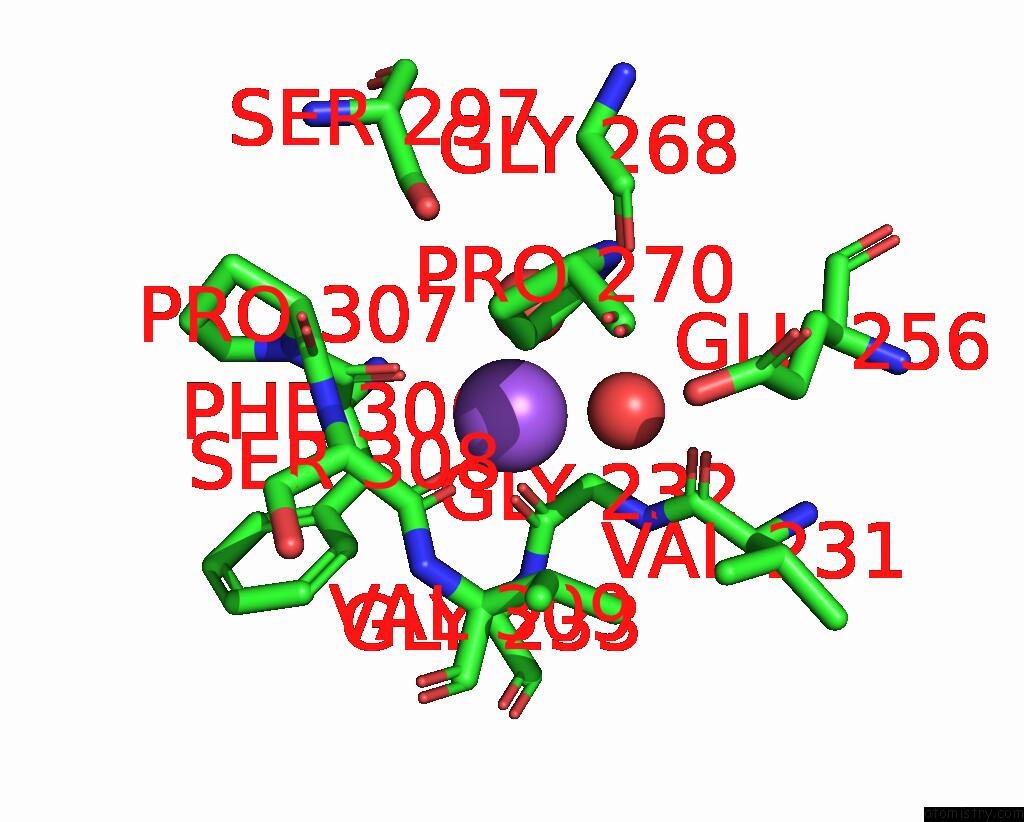
Mono view
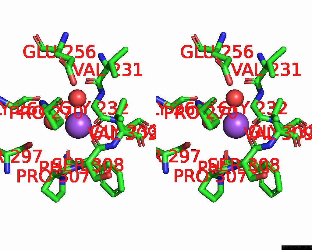
Stereo pair view
Mono view
Stereo pair view
A full contact list of Sodium with other atoms in the Na binding
site number 1 of Crystal Structure of Wild-Type Tryptophan Synthase Complexed with N- [1H-Indol-3-Yl-Acetyl]Glycine Acid within 5.0Å range:
|
Reference:
M.Weyand,
I.Schlichting,
A.Marabotti,
A.Mozzarelli.
Crystal Structures of A New Class of Allosteric Effectors Complexed to Tryptophan Synthase. J.Biol.Chem. V. 277 10647 2002.
ISSN: ISSN 0021-9258
PubMed: 11756456
DOI: 10.1074/JBC.M111285200
Page generated: Sun Aug 17 05:45:26 2025
ISSN: ISSN 0021-9258
PubMed: 11756456
DOI: 10.1074/JBC.M111285200
Last articles
Na in 2H34Na in 2GW0
Na in 2H4V
Na in 2GYQ
Na in 2GXB
Na in 2GUM
Na in 2GUO
Na in 2GVH
Na in 2GVK
Na in 2GU7