Sodium »
PDB 3e40-3ept »
3epi »
Sodium in PDB 3epi: Structure of Human Dna Polymerase Iota Complexed with N2-Ethylguanine and Incoming Ttp
Enzymatic activity of Structure of Human Dna Polymerase Iota Complexed with N2-Ethylguanine and Incoming Ttp
All present enzymatic activity of Structure of Human Dna Polymerase Iota Complexed with N2-Ethylguanine and Incoming Ttp:
2.7.7.7;
2.7.7.7;
Protein crystallography data
The structure of Structure of Human Dna Polymerase Iota Complexed with N2-Ethylguanine and Incoming Ttp, PDB code: 3epi
was solved by
M.G.Pence,
with X-Ray Crystallography technique. A brief refinement statistics is given in the table below:
| Resolution Low / High (Å) | 14.99 / 2.90 |
| Space group | P 65 2 2 |
| Cell size a, b, c (Å), α, β, γ (°) | 98.643, 98.643, 202.231, 90.00, 90.00, 120.00 |
| R / Rfree (%) | 23.6 / 28.2 |
Sodium Binding Sites:
The binding sites of Sodium atom in the Structure of Human Dna Polymerase Iota Complexed with N2-Ethylguanine and Incoming Ttp
(pdb code 3epi). This binding sites where shown within
5.0 Angstroms radius around Sodium atom.
In total only one binding site of Sodium was determined in the Structure of Human Dna Polymerase Iota Complexed with N2-Ethylguanine and Incoming Ttp, PDB code: 3epi:
In total only one binding site of Sodium was determined in the Structure of Human Dna Polymerase Iota Complexed with N2-Ethylguanine and Incoming Ttp, PDB code: 3epi:
Sodium binding site 1 out of 1 in 3epi
Go back to
Sodium binding site 1 out
of 1 in the Structure of Human Dna Polymerase Iota Complexed with N2-Ethylguanine and Incoming Ttp
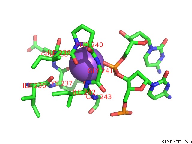
Mono view
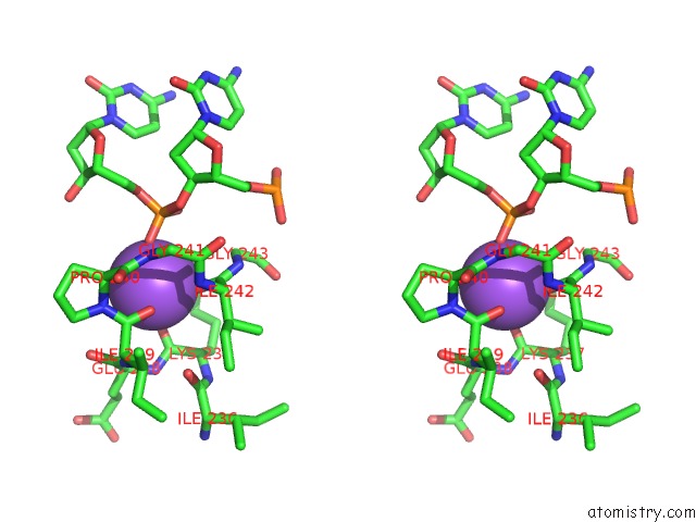
Stereo pair view
Mono view
Stereo pair view
A full contact list of Sodium with other atoms in the Na binding
site number 1 of Structure of Human Dna Polymerase Iota Complexed with N2-Ethylguanine and Incoming Ttp within 5.0Å range:
|
Reference:
M.G.Pence,
P.Blans,
C.N.Zink,
T.Hollis,
J.C.Fishbein,
F.W.Perrino.
Lesion Bypass of N2-Ethylguanine By Human Dna Polymerase Iota. J.Biol.Chem. V. 284 1732 2009.
ISSN: ISSN 0021-9258
PubMed: 18984581
DOI: 10.1074/JBC.M807296200
Page generated: Sun Aug 17 14:43:06 2025
ISSN: ISSN 0021-9258
PubMed: 18984581
DOI: 10.1074/JBC.M807296200
Last articles
Na in 5EK5Na in 5EHW
Na in 5EFO
Na in 5EHV
Na in 5EH7
Na in 5EGM
Na in 5EE5
Na in 5EG2
Na in 5E9S
Na in 5EFI