Sodium »
PDB 5dyn-5emg »
5efo »
Sodium in PDB 5efo: X-Ray Structure Uridine Phosphorylase From Vibrio Cholerae in Complex with Cytidine and Cytosine at 1.63A.
Enzymatic activity of X-Ray Structure Uridine Phosphorylase From Vibrio Cholerae in Complex with Cytidine and Cytosine at 1.63A.
All present enzymatic activity of X-Ray Structure Uridine Phosphorylase From Vibrio Cholerae in Complex with Cytidine and Cytosine at 1.63A.:
2.4.2.3;
2.4.2.3;
Protein crystallography data
The structure of X-Ray Structure Uridine Phosphorylase From Vibrio Cholerae in Complex with Cytidine and Cytosine at 1.63A., PDB code: 5efo
was solved by
I.I.Prokofev,
A.A.Lashkov,
A.G.Gabdoulkhakov,
C.Betzel,
A.M.Mikhailov,
with X-Ray Crystallography technique. A brief refinement statistics is given in the table below:
| Resolution Low / High (Å) | 19.97 / 1.63 |
| Space group | P 1 |
| Cell size a, b, c (Å), α, β, γ (°) | 59.949, 76.336, 89.694, 67.54, 73.74, 84.92 |
| R / Rfree (%) | 16.3 / 20.7 |
Sodium Binding Sites:
The binding sites of Sodium atom in the X-Ray Structure Uridine Phosphorylase From Vibrio Cholerae in Complex with Cytidine and Cytosine at 1.63A.
(pdb code 5efo). This binding sites where shown within
5.0 Angstroms radius around Sodium atom.
In total 3 binding sites of Sodium where determined in the X-Ray Structure Uridine Phosphorylase From Vibrio Cholerae in Complex with Cytidine and Cytosine at 1.63A., PDB code: 5efo:
Jump to Sodium binding site number: 1; 2; 3;
In total 3 binding sites of Sodium where determined in the X-Ray Structure Uridine Phosphorylase From Vibrio Cholerae in Complex with Cytidine and Cytosine at 1.63A., PDB code: 5efo:
Jump to Sodium binding site number: 1; 2; 3;
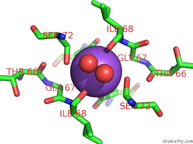
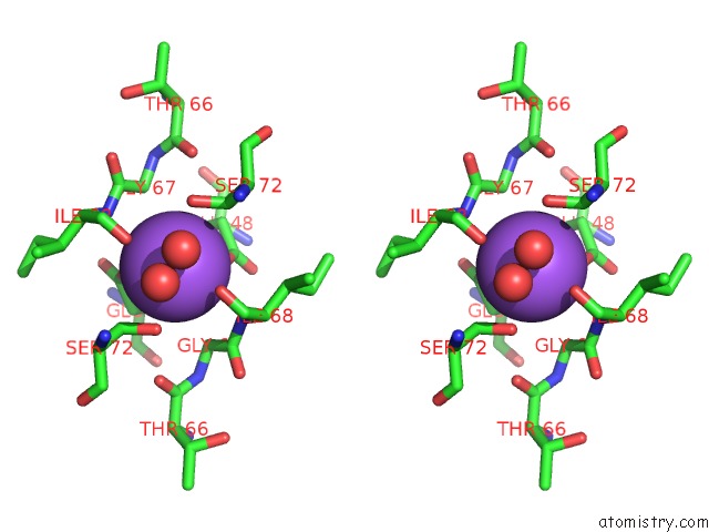
Sodium binding site 1 out of 3 in 5efo
Go back to
Sodium binding site 1 out
of 3 in the X-Ray Structure Uridine Phosphorylase From Vibrio Cholerae in Complex with Cytidine and Cytosine at 1.63A.
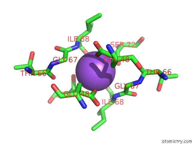
Mono view
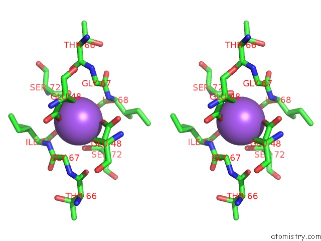
Stereo pair view
Mono view
Stereo pair view
A full contact list of Sodium with other atoms in the Na binding
site number 1 of X-Ray Structure Uridine Phosphorylase From Vibrio Cholerae in Complex with Cytidine and Cytosine at 1.63A. within 5.0Å range:
|
Sodium binding site 2 out of 3 in 5efo
Go back to
Sodium binding site 2 out
of 3 in the X-Ray Structure Uridine Phosphorylase From Vibrio Cholerae in Complex with Cytidine and Cytosine at 1.63A.
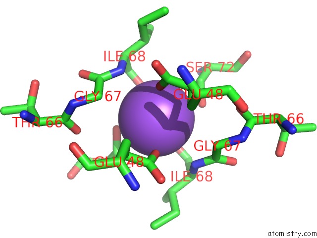
Mono view
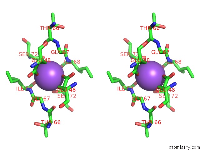
Stereo pair view
Mono view
Stereo pair view
A full contact list of Sodium with other atoms in the Na binding
site number 2 of X-Ray Structure Uridine Phosphorylase From Vibrio Cholerae in Complex with Cytidine and Cytosine at 1.63A. within 5.0Å range:
|
Sodium binding site 3 out of 3 in 5efo
Go back to
Sodium binding site 3 out
of 3 in the X-Ray Structure Uridine Phosphorylase From Vibrio Cholerae in Complex with Cytidine and Cytosine at 1.63A.
Mono view
Stereo pair view
Mono view
Stereo pair view
A full contact list of Sodium with other atoms in the Na binding
site number 3 of X-Ray Structure Uridine Phosphorylase From Vibrio Cholerae in Complex with Cytidine and Cytosine at 1.63A. within 5.0Å range:
|
Reference:
I.I.Prokofev,
A.A.Lashkov,
A.G.Gabdoulkhakov,
C.Betzel,
A.M.Mikhailov.
X-Ray Structure Uridine Phosphorylase From Vibrio Cholerae in Complex with Uridine at 2.24 A Resolution To Be Published.
Page generated: Sun Aug 17 23:37:50 2025
Last articles
Zn in 4GK8Zn in 4GIZ
Zn in 4GKK
Zn in 4GKJ
Zn in 4GIY
Zn in 4GI4
Zn in 4GH6
Zn in 4GHR
Zn in 4GH3
Zn in 4GH1