Sodium »
PDB 3cq8-3dfh »
3d97 »
Sodium in PDB 3d97: Crystal Structure of the R132K:R111L:L121E Mutant of Apo-Cellular Retinoic Acid Binding Protein Type II at 1.50 Angstroms Resolution
Protein crystallography data
The structure of Crystal Structure of the R132K:R111L:L121E Mutant of Apo-Cellular Retinoic Acid Binding Protein Type II at 1.50 Angstroms Resolution, PDB code: 3d97
was solved by
S.Vaezeslami,
J.H.Geiger,
with X-Ray Crystallography technique. A brief refinement statistics is given in the table below:
| Resolution Low / High (Å) | 55.90 / 1.50 |
| Space group | P 1 |
| Cell size a, b, c (Å), α, β, γ (°) | 34.553, 37.220, 61.058, 73.40, 73.70, 89.73 |
| R / Rfree (%) | 15.4 / 22.7 |
Sodium Binding Sites:
The binding sites of Sodium atom in the Crystal Structure of the R132K:R111L:L121E Mutant of Apo-Cellular Retinoic Acid Binding Protein Type II at 1.50 Angstroms Resolution
(pdb code 3d97). This binding sites where shown within
5.0 Angstroms radius around Sodium atom.
In total 2 binding sites of Sodium where determined in the Crystal Structure of the R132K:R111L:L121E Mutant of Apo-Cellular Retinoic Acid Binding Protein Type II at 1.50 Angstroms Resolution, PDB code: 3d97:
Jump to Sodium binding site number: 1; 2;
In total 2 binding sites of Sodium where determined in the Crystal Structure of the R132K:R111L:L121E Mutant of Apo-Cellular Retinoic Acid Binding Protein Type II at 1.50 Angstroms Resolution, PDB code: 3d97:
Jump to Sodium binding site number: 1; 2;
Sodium binding site 1 out of 2 in 3d97
Go back to
Sodium binding site 1 out
of 2 in the Crystal Structure of the R132K:R111L:L121E Mutant of Apo-Cellular Retinoic Acid Binding Protein Type II at 1.50 Angstroms Resolution
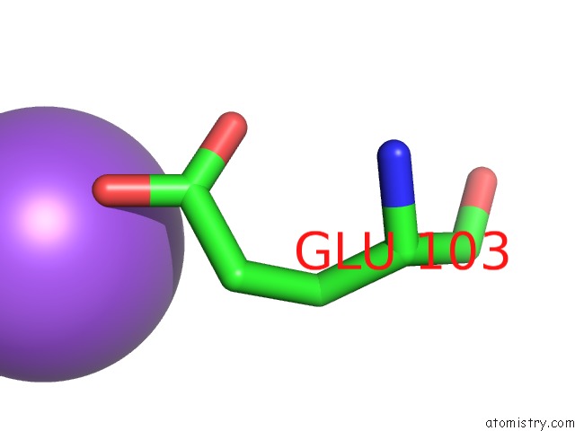
Mono view
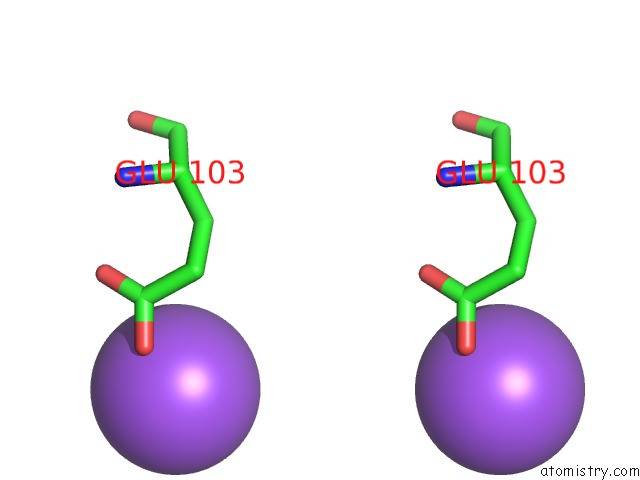
Stereo pair view
Mono view
Stereo pair view
A full contact list of Sodium with other atoms in the Na binding
site number 1 of Crystal Structure of the R132K:R111L:L121E Mutant of Apo-Cellular Retinoic Acid Binding Protein Type II at 1.50 Angstroms Resolution within 5.0Å range:
|
Sodium binding site 2 out of 2 in 3d97
Go back to
Sodium binding site 2 out
of 2 in the Crystal Structure of the R132K:R111L:L121E Mutant of Apo-Cellular Retinoic Acid Binding Protein Type II at 1.50 Angstroms Resolution
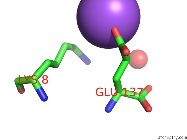
Mono view
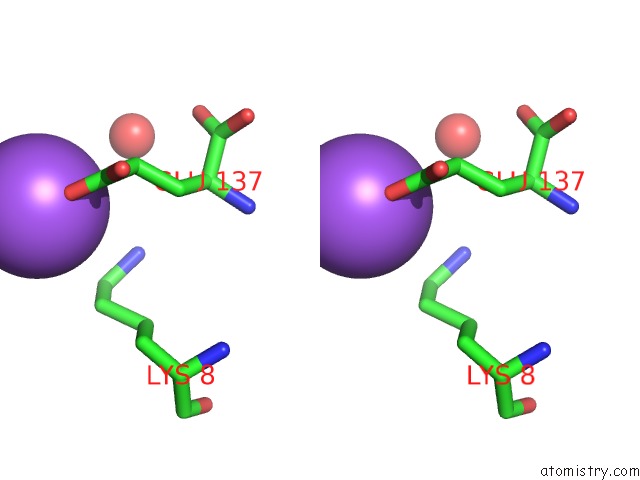
Stereo pair view
Mono view
Stereo pair view
A full contact list of Sodium with other atoms in the Na binding
site number 2 of Crystal Structure of the R132K:R111L:L121E Mutant of Apo-Cellular Retinoic Acid Binding Protein Type II at 1.50 Angstroms Resolution within 5.0Å range:
|
Reference:
S.Vaezeslami,
X.Jia,
C.Vasileiou,
B.Borhan,
J.H.Geiger.
Determining Crystal Structures of Proteins and Protein Complexes By X-Ray Crystallography: X-Ray Crystallographic Studies of the Mutants of Cellular Retinoic Acid Binding Protein Type II Toward Designing A Mimic of Rhodopsin. Thesis.
Page generated: Sun Aug 17 14:19:14 2025
Last articles
Na in 6J8ENa in 6J6V
Na in 6J6O
Na in 6J3B
Na in 6J05
Na in 6J0H
Na in 6IWH
Na in 6IX8
Na in 6IX9
Na in 6IWG