Sodium »
PDB 9c42-9dpo »
9cum »
Sodium in PDB 9cum: Q67H Mutant of R67 Dhfr Complexed with Congo Red
Enzymatic activity of Q67H Mutant of R67 Dhfr Complexed with Congo Red
All present enzymatic activity of Q67H Mutant of R67 Dhfr Complexed with Congo Red:
1.5.1.3;
1.5.1.3;
Protein crystallography data
The structure of Q67H Mutant of R67 Dhfr Complexed with Congo Red, PDB code: 9cum
was solved by
N.Narayana,
A.N.Narendra,
with X-Ray Crystallography technique. A brief refinement statistics is given in the table below:
| Resolution Low / High (Å) | 15.00 / 1.15 |
| Space group | I 41 2 2 |
| Cell size a, b, c (Å), α, β, γ (°) | 67.303, 67.303, 52.684, 90, 90, 90 |
| R / Rfree (%) | 12.2 / 13.5 |
Sodium Binding Sites:
The binding sites of Sodium atom in the Q67H Mutant of R67 Dhfr Complexed with Congo Red
(pdb code 9cum). This binding sites where shown within
5.0 Angstroms radius around Sodium atom.
In total only one binding site of Sodium was determined in the Q67H Mutant of R67 Dhfr Complexed with Congo Red, PDB code: 9cum:
In total only one binding site of Sodium was determined in the Q67H Mutant of R67 Dhfr Complexed with Congo Red, PDB code: 9cum:
Sodium binding site 1 out of 1 in 9cum
Go back to
Sodium binding site 1 out
of 1 in the Q67H Mutant of R67 Dhfr Complexed with Congo Red
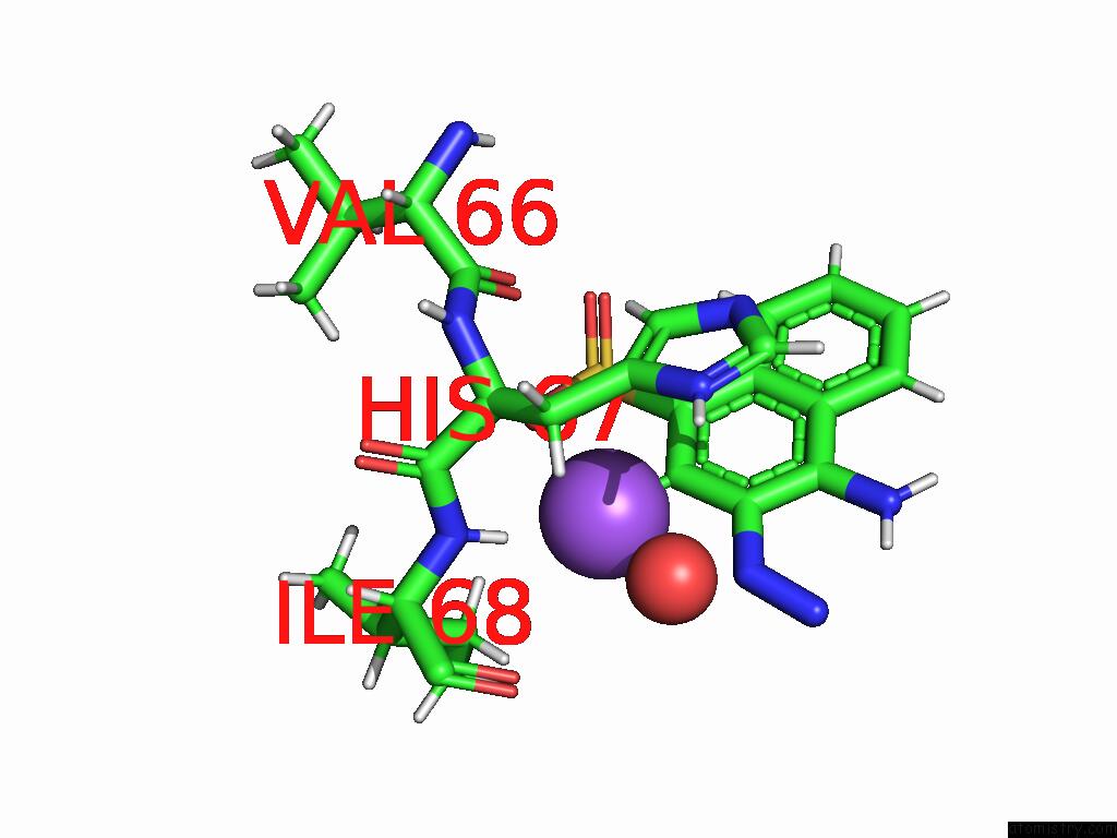
Mono view
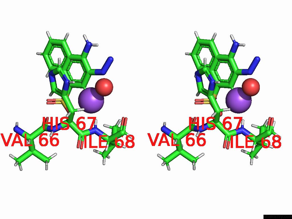
Stereo pair view
Mono view
Stereo pair view
A full contact list of Sodium with other atoms in the Na binding
site number 1 of Q67H Mutant of R67 Dhfr Complexed with Congo Red within 5.0Å range:
|
Reference:
A.N.Narendra,
E.E.Howell,
N.Narayana.
Crystal Structure of the Plasmid-Encoded R67 Dihydrofolate Reductase Complexed with Congo Red An Amyloid Binding Dye Sci Rep V. 15 5212 2025.
ISSN: ESSN 2045-2322
DOI: 10.1038/S41598-025-89539-3
Page generated: Mon Aug 18 16:39:18 2025
ISSN: ESSN 2045-2322
DOI: 10.1038/S41598-025-89539-3
Last articles
Mn in 9LJUMn in 9LJW
Mn in 9LJS
Mn in 9LJR
Mn in 9LJT
Mn in 9LJV
Mg in 9UA2
Mg in 9R96
Mg in 9VM1
Mg in 9P01