Sodium »
PDB 8vnr-8wch »
8w8v »
Sodium in PDB 8w8v: High-Resolution X-Ray Structure of Cellulase CEL6A From Phanerochaete Chrysosporium at Cryogenic Temperature, Enzyme-Product Complex
Protein crystallography data
The structure of High-Resolution X-Ray Structure of Cellulase CEL6A From Phanerochaete Chrysosporium at Cryogenic Temperature, Enzyme-Product Complex, PDB code: 8w8v
was solved by
M.Tachioka,
S.Yamaguchi,
A.Nakamura,
T.Ishida,
K.Kusaka,
T.Yamada,
N.Yano,
T.Chatake,
T.Tamada,
K.Takeda,
S.Niwa,
H.Tanaka,
S.Takahashi,
K.Inaka,
N.Furubayashi,
S.Deguchi,
M.Samejima,
K.Igarashi,
with X-Ray Crystallography technique. A brief refinement statistics is given in the table below:
| Resolution Low / High (Å) | 46.10 / 0.85 |
| Space group | P 21 21 21 |
| Cell size a, b, c (Å), α, β, γ (°) | 54.044, 67.581, 88.354, 90, 90, 90 |
| R / Rfree (%) | n/a / n/a |
Other elements in 8w8v:
The structure of High-Resolution X-Ray Structure of Cellulase CEL6A From Phanerochaete Chrysosporium at Cryogenic Temperature, Enzyme-Product Complex also contains other interesting chemical elements:
| Chlorine | (Cl) | 1 atom |
Sodium Binding Sites:
The binding sites of Sodium atom in the High-Resolution X-Ray Structure of Cellulase CEL6A From Phanerochaete Chrysosporium at Cryogenic Temperature, Enzyme-Product Complex
(pdb code 8w8v). This binding sites where shown within
5.0 Angstroms radius around Sodium atom.
In total only one binding site of Sodium was determined in the High-Resolution X-Ray Structure of Cellulase CEL6A From Phanerochaete Chrysosporium at Cryogenic Temperature, Enzyme-Product Complex, PDB code: 8w8v:
In total only one binding site of Sodium was determined in the High-Resolution X-Ray Structure of Cellulase CEL6A From Phanerochaete Chrysosporium at Cryogenic Temperature, Enzyme-Product Complex, PDB code: 8w8v:
Sodium binding site 1 out of 1 in 8w8v
Go back to
Sodium binding site 1 out
of 1 in the High-Resolution X-Ray Structure of Cellulase CEL6A From Phanerochaete Chrysosporium at Cryogenic Temperature, Enzyme-Product Complex
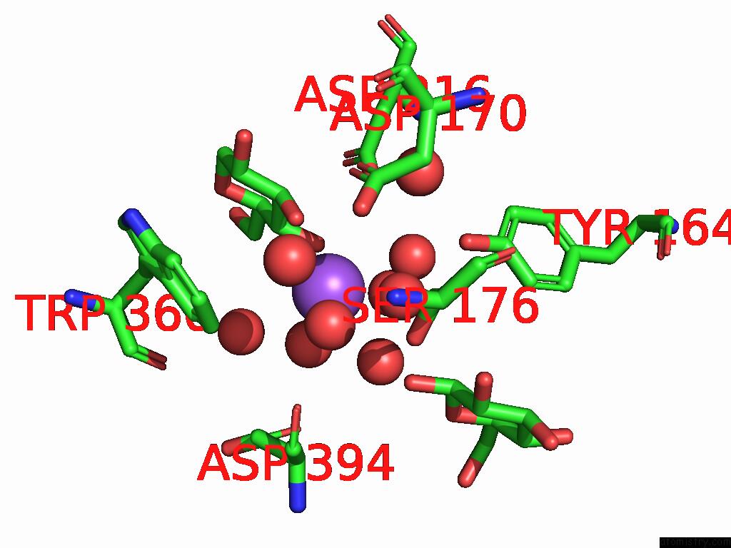
Mono view
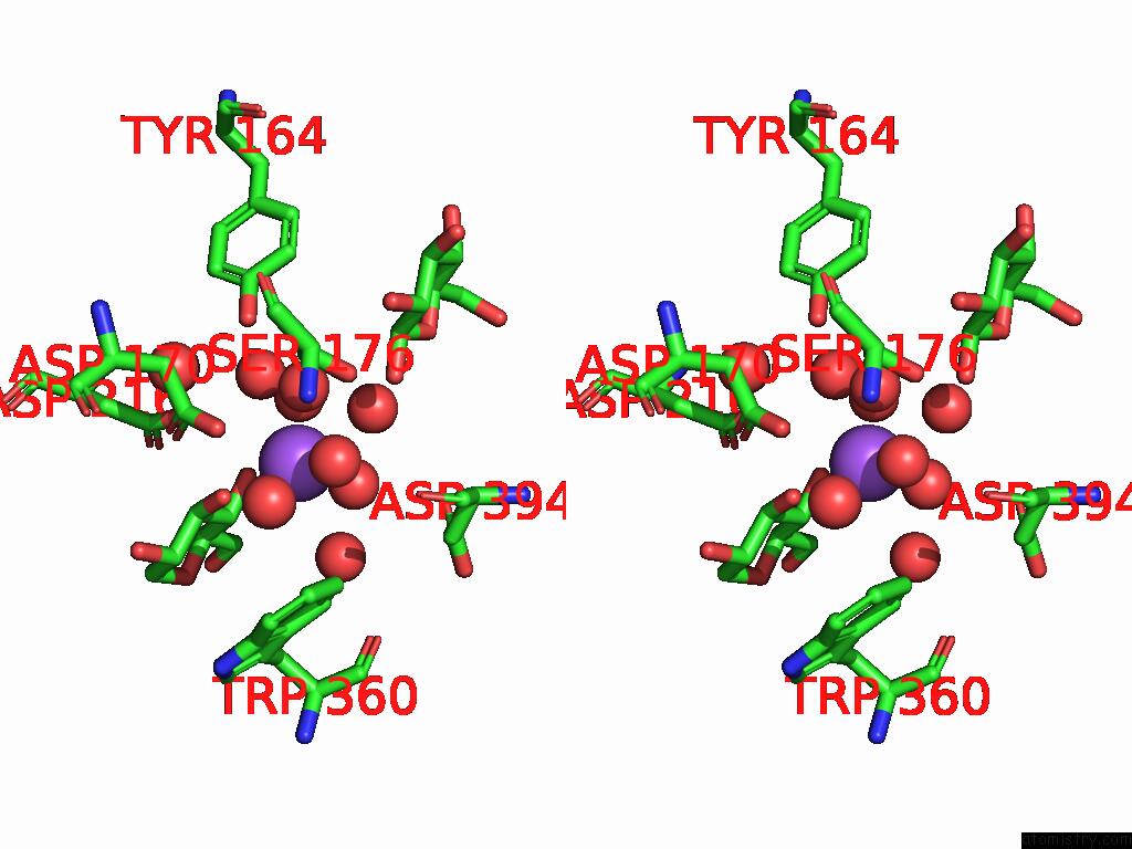
Stereo pair view
Mono view
Stereo pair view
A full contact list of Sodium with other atoms in the Na binding
site number 1 of High-Resolution X-Ray Structure of Cellulase CEL6A From Phanerochaete Chrysosporium at Cryogenic Temperature, Enzyme-Product Complex within 5.0Å range:
|
Reference:
M.Tachioka,
S.Yamaguchi,
A.Nakamura,
T.Ishida,
K.Kusaka,
T.Yamada,
N.Yano,
T.Chatake,
T.Tamada,
K.Takeda,
S.Niwa,
H.Tanaka,
S.Takahashi,
K.Inaka,
N.Furubayashi,
S.Deguchi,
M.Samejima,
K.Igarashi.
Deprotonated Arginine Controls A Putative Catalytic Base in Invert-Ing Family 6 Glycoside Hydrolase To Be Published.
Page generated: Mon Aug 18 16:16:37 2025
Last articles
K in 9NESK in 9PHG
K in 9NEI
K in 9NED
K in 9NEC
K in 9NEG
K in 9CWU
K in 9CVB
K in 9CVA
K in 9COM