Sodium »
PDB 7e4c-7f6s »
7esk »
Sodium in PDB 7esk: Crystal Structure of A L-Rhamnose-Alpha-1,4-D-Glucuronate Lyase From Fusarium Oxysporum 12S, Ligand Free Form
Protein crystallography data
The structure of Crystal Structure of A L-Rhamnose-Alpha-1,4-D-Glucuronate Lyase From Fusarium Oxysporum 12S, Ligand Free Form, PDB code: 7esk
was solved by
T.Kondo,
T.Arakawa,
S.Fushinobu,
T.Sakamoto,
with X-Ray Crystallography technique. A brief refinement statistics is given in the table below:
| Resolution Low / High (Å) | 31.30 / 1.05 |
| Space group | P 21 21 21 |
| Cell size a, b, c (Å), α, β, γ (°) | 58.113, 65.267, 107.008, 90, 90, 90 |
| R / Rfree (%) | 15.1 / 17.1 |
Other elements in 7esk:
The structure of Crystal Structure of A L-Rhamnose-Alpha-1,4-D-Glucuronate Lyase From Fusarium Oxysporum 12S, Ligand Free Form also contains other interesting chemical elements:
| Calcium | (Ca) | 1 atom |
Sodium Binding Sites:
The binding sites of Sodium atom in the Crystal Structure of A L-Rhamnose-Alpha-1,4-D-Glucuronate Lyase From Fusarium Oxysporum 12S, Ligand Free Form
(pdb code 7esk). This binding sites where shown within
5.0 Angstroms radius around Sodium atom.
In total only one binding site of Sodium was determined in the Crystal Structure of A L-Rhamnose-Alpha-1,4-D-Glucuronate Lyase From Fusarium Oxysporum 12S, Ligand Free Form, PDB code: 7esk:
In total only one binding site of Sodium was determined in the Crystal Structure of A L-Rhamnose-Alpha-1,4-D-Glucuronate Lyase From Fusarium Oxysporum 12S, Ligand Free Form, PDB code: 7esk:
Sodium binding site 1 out of 1 in 7esk
Go back to
Sodium binding site 1 out
of 1 in the Crystal Structure of A L-Rhamnose-Alpha-1,4-D-Glucuronate Lyase From Fusarium Oxysporum 12S, Ligand Free Form
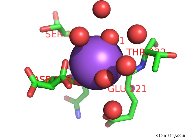
Mono view
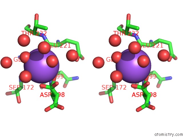
Stereo pair view
Mono view
Stereo pair view
A full contact list of Sodium with other atoms in the Na binding
site number 1 of Crystal Structure of A L-Rhamnose-Alpha-1,4-D-Glucuronate Lyase From Fusarium Oxysporum 12S, Ligand Free Form within 5.0Å range:
|
Reference:
T.Kondo,
M.Kichijo,
A.Maruta,
M.Nakaya,
S.Takenaka,
T.Arakawa,
S.Fushinobu,
T.Sakamoto.
Structural and Functional Analysis of Gum Arabic L-Rhamnose-Alpha-1,4-D-Glucuronate Lyase Establishes A Novel Polysaccharide Lyase Family. J.Biol.Chem. 01001 2021.
ISSN: ESSN 1083-351X
PubMed: 34303708
DOI: 10.1016/J.JBC.2021.101001
Page generated: Mon Aug 18 09:43:23 2025
ISSN: ESSN 1083-351X
PubMed: 34303708
DOI: 10.1016/J.JBC.2021.101001
Last articles
Mn in 9LJUMn in 9LJW
Mn in 9LJS
Mn in 9LJR
Mn in 9LJT
Mn in 9LJV
Mg in 9UA2
Mg in 9R96
Mg in 9VM1
Mg in 9P01