Sodium »
PDB 6s08-6sez »
6s3b »
Sodium in PDB 6s3b: Ligand Binding Domain of the P. Putida Receptor PCAY_PP in Complex with Benzoate
Protein crystallography data
The structure of Ligand Binding Domain of the P. Putida Receptor PCAY_PP in Complex with Benzoate, PDB code: 6s3b
was solved by
J.A.Gavira,
M.A.Mantilla,
M.Fernandez,
T.Krell,
with X-Ray Crystallography technique. A brief refinement statistics is given in the table below:
| Resolution Low / High (Å) | 46.17 / 1.95 |
| Space group | P 21 21 21 |
| Cell size a, b, c (Å), α, β, γ (°) | 45.390, 69.219, 92.333, 90.00, 90.00, 90.00 |
| R / Rfree (%) | 17.6 / 21.6 |
Sodium Binding Sites:
The binding sites of Sodium atom in the Ligand Binding Domain of the P. Putida Receptor PCAY_PP in Complex with Benzoate
(pdb code 6s3b). This binding sites where shown within
5.0 Angstroms radius around Sodium atom.
In total only one binding site of Sodium was determined in the Ligand Binding Domain of the P. Putida Receptor PCAY_PP in Complex with Benzoate, PDB code: 6s3b:
In total only one binding site of Sodium was determined in the Ligand Binding Domain of the P. Putida Receptor PCAY_PP in Complex with Benzoate, PDB code: 6s3b:
Sodium binding site 1 out of 1 in 6s3b
Go back to
Sodium binding site 1 out
of 1 in the Ligand Binding Domain of the P. Putida Receptor PCAY_PP in Complex with Benzoate
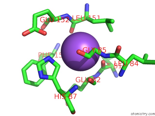
Mono view
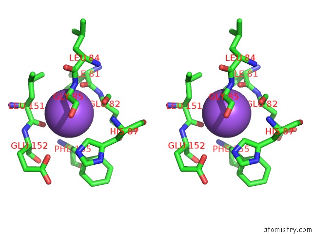
Stereo pair view
Mono view
Stereo pair view
A full contact list of Sodium with other atoms in the Na binding
site number 1 of Ligand Binding Domain of the P. Putida Receptor PCAY_PP in Complex with Benzoate within 5.0Å range:
|
Reference:
J.A.Gavira,
M.A.Matilla,
M.Fernandez,
T.Krell.
The Structural Basis For Signal Promiscuity in A Bacterial Chemoreceptor. Febs J. 2020.
ISSN: ISSN 1742-464X
PubMed: 33021055
DOI: 10.1111/FEBS.15580
Page generated: Tue Oct 8 13:22:18 2024
ISSN: ISSN 1742-464X
PubMed: 33021055
DOI: 10.1111/FEBS.15580
Last articles
Fe in 2YXOFe in 2YRS
Fe in 2YXC
Fe in 2YNM
Fe in 2YVJ
Fe in 2YP1
Fe in 2YU2
Fe in 2YU1
Fe in 2YQB
Fe in 2YOO