Sodium »
PDB 5cwy-5ddp »
5d18 »
Sodium in PDB 5d18: Crystal Structure of Mycobacterium Tuberculosis RV0302, Form I
Protein crystallography data
The structure of Crystal Structure of Mycobacterium Tuberculosis RV0302, Form I, PDB code: 5d18
was solved by
T.-H.Chou,
J.Delmar,
C.-C.Su,
E.Yu,
with X-Ray Crystallography technique. A brief refinement statistics is given in the table below:
| Resolution Low / High (Å) | 38.18 / 2.04 |
| Space group | P 61 2 2 |
| Cell size a, b, c (Å), α, β, γ (°) | 116.628, 116.628, 94.178, 90.00, 90.00, 120.00 |
| R / Rfree (%) | 19.2 / 21.5 |
Sodium Binding Sites:
The binding sites of Sodium atom in the Crystal Structure of Mycobacterium Tuberculosis RV0302, Form I
(pdb code 5d18). This binding sites where shown within
5.0 Angstroms radius around Sodium atom.
In total only one binding site of Sodium was determined in the Crystal Structure of Mycobacterium Tuberculosis RV0302, Form I, PDB code: 5d18:
In total only one binding site of Sodium was determined in the Crystal Structure of Mycobacterium Tuberculosis RV0302, Form I, PDB code: 5d18:
Sodium binding site 1 out of 1 in 5d18
Go back to
Sodium binding site 1 out
of 1 in the Crystal Structure of Mycobacterium Tuberculosis RV0302, Form I
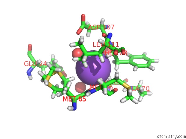
Mono view
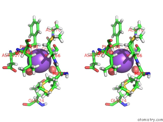
Stereo pair view
Mono view
Stereo pair view
A full contact list of Sodium with other atoms in the Na binding
site number 1 of Crystal Structure of Mycobacterium Tuberculosis RV0302, Form I within 5.0Å range:
|
Reference:
T.H.Chou,
J.A.Delmar,
C.C.Wright,
N.Kumar,
A.Radhakrishnan,
J.K.Doh,
M.H.Licon,
J.Reddy Bolla,
H.T.Lei,
K.R.Rajashankar,
C.C.Su,
G.E.Purdy,
E.W.Yu.
Crystal Structure of the Mycobacterium Tuberculosis Transcriptional Regulator RV0302. Protein Sci. 2015.
ISSN: ESSN 1469-896X
PubMed: 26362239
DOI: 10.1002/PRO.2802
Page generated: Mon Oct 7 20:28:30 2024
ISSN: ESSN 1469-896X
PubMed: 26362239
DOI: 10.1002/PRO.2802
Last articles
K in 7NPXK in 7NWD
K in 7NXF
K in 7NF4
K in 7NHT
K in 7NKG
K in 7NF2
K in 7N9L
K in 7NFZ
K in 7NF3