Sodium »
PDB 4ppf-4q3c »
4pvr »
Sodium in PDB 4pvr: Crystal Structure of Partially-Cleaved Human L-Asparaginase Protein in Complex with L-Aspartate
Enzymatic activity of Crystal Structure of Partially-Cleaved Human L-Asparaginase Protein in Complex with L-Aspartate
All present enzymatic activity of Crystal Structure of Partially-Cleaved Human L-Asparaginase Protein in Complex with L-Aspartate:
3.4.19.5; 3.5.1.1;
3.4.19.5; 3.5.1.1;
Protein crystallography data
The structure of Crystal Structure of Partially-Cleaved Human L-Asparaginase Protein in Complex with L-Aspartate, PDB code: 4pvr
was solved by
J.Nomme,
A.Lavie,
with X-Ray Crystallography technique. A brief refinement statistics is given in the table below:
| Resolution Low / High (Å) | 30.00 / 1.75 |
| Space group | P 65 |
| Cell size a, b, c (Å), α, β, γ (°) | 59.500, 59.500, 299.500, 90.00, 90.00, 120.00 |
| R / Rfree (%) | 15.9 / 19.1 |
Sodium Binding Sites:
The binding sites of Sodium atom in the Crystal Structure of Partially-Cleaved Human L-Asparaginase Protein in Complex with L-Aspartate
(pdb code 4pvr). This binding sites where shown within
5.0 Angstroms radius around Sodium atom.
In total 2 binding sites of Sodium where determined in the Crystal Structure of Partially-Cleaved Human L-Asparaginase Protein in Complex with L-Aspartate, PDB code: 4pvr:
Jump to Sodium binding site number: 1; 2;
In total 2 binding sites of Sodium where determined in the Crystal Structure of Partially-Cleaved Human L-Asparaginase Protein in Complex with L-Aspartate, PDB code: 4pvr:
Jump to Sodium binding site number: 1; 2;
Sodium binding site 1 out of 2 in 4pvr
Go back to
Sodium binding site 1 out
of 2 in the Crystal Structure of Partially-Cleaved Human L-Asparaginase Protein in Complex with L-Aspartate
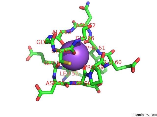
Mono view
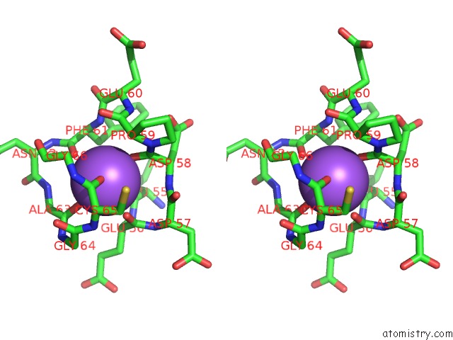
Stereo pair view
Mono view
Stereo pair view
A full contact list of Sodium with other atoms in the Na binding
site number 1 of Crystal Structure of Partially-Cleaved Human L-Asparaginase Protein in Complex with L-Aspartate within 5.0Å range:
|
Sodium binding site 2 out of 2 in 4pvr
Go back to
Sodium binding site 2 out
of 2 in the Crystal Structure of Partially-Cleaved Human L-Asparaginase Protein in Complex with L-Aspartate
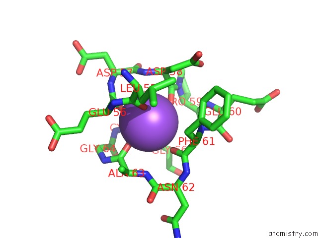
Mono view
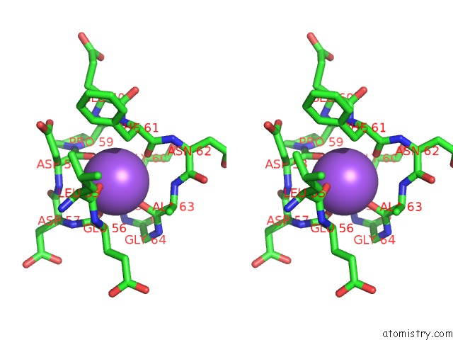
Stereo pair view
Mono view
Stereo pair view
A full contact list of Sodium with other atoms in the Na binding
site number 2 of Crystal Structure of Partially-Cleaved Human L-Asparaginase Protein in Complex with L-Aspartate within 5.0Å range:
|
Reference:
J.Nomme,
Y.Su,
M.Konrad,
A.Lavie.
Structures of Apo and Product-Bound Human L-Asparaginase: Insights Into the Mechanism of Autoproteolysis and Substrate Hydrolysis. Biochemistry V. 51 6816 2012.
ISSN: ISSN 0006-2960
PubMed: 22861376
DOI: 10.1021/BI300870G
Page generated: Sun Aug 17 21:17:45 2025
ISSN: ISSN 0006-2960
PubMed: 22861376
DOI: 10.1021/BI300870G
Last articles
Na in 5MLRNa in 5MLS
Na in 5MIW
Na in 5MJT
Na in 5MLH
Na in 5MK6
Na in 5MJJ
Na in 5MIY
Na in 5MJG
Na in 5MIM