Sodium »
PDB 4m04-4mfa »
4ma5 »
Sodium in PDB 4ma5: The Crystal Structure of Phosphoribosylaminoimidazole Carboxylase Atpase Subunit of Francisella Tularensis Subsp. Tularensis Schu S4 in Complex with An Atp Analog, Amp-Pnp.
Enzymatic activity of The Crystal Structure of Phosphoribosylaminoimidazole Carboxylase Atpase Subunit of Francisella Tularensis Subsp. Tularensis Schu S4 in Complex with An Atp Analog, Amp-Pnp.
All present enzymatic activity of The Crystal Structure of Phosphoribosylaminoimidazole Carboxylase Atpase Subunit of Francisella Tularensis Subsp. Tularensis Schu S4 in Complex with An Atp Analog, Amp-Pnp.:
4.1.1.21;
4.1.1.21;
Protein crystallography data
The structure of The Crystal Structure of Phosphoribosylaminoimidazole Carboxylase Atpase Subunit of Francisella Tularensis Subsp. Tularensis Schu S4 in Complex with An Atp Analog, Amp-Pnp., PDB code: 4ma5
was solved by
K.Tan,
M.Zhou,
K.Kwon,
W.F.Anderson,
A.Joachimiak,
Center For Structuralgenomics Of Infectious Diseases (Csgid),
with X-Ray Crystallography technique. A brief refinement statistics is given in the table below:
| Resolution Low / High (Å) | 26.86 / 1.81 |
| Space group | P 43 21 2 |
| Cell size a, b, c (Å), α, β, γ (°) | 84.943, 84.943, 106.539, 90.00, 90.00, 90.00 |
| R / Rfree (%) | 17.3 / 21.6 |
Sodium Binding Sites:
The binding sites of Sodium atom in the The Crystal Structure of Phosphoribosylaminoimidazole Carboxylase Atpase Subunit of Francisella Tularensis Subsp. Tularensis Schu S4 in Complex with An Atp Analog, Amp-Pnp.
(pdb code 4ma5). This binding sites where shown within
5.0 Angstroms radius around Sodium atom.
In total 2 binding sites of Sodium where determined in the The Crystal Structure of Phosphoribosylaminoimidazole Carboxylase Atpase Subunit of Francisella Tularensis Subsp. Tularensis Schu S4 in Complex with An Atp Analog, Amp-Pnp., PDB code: 4ma5:
Jump to Sodium binding site number: 1; 2;
In total 2 binding sites of Sodium where determined in the The Crystal Structure of Phosphoribosylaminoimidazole Carboxylase Atpase Subunit of Francisella Tularensis Subsp. Tularensis Schu S4 in Complex with An Atp Analog, Amp-Pnp., PDB code: 4ma5:
Jump to Sodium binding site number: 1; 2;
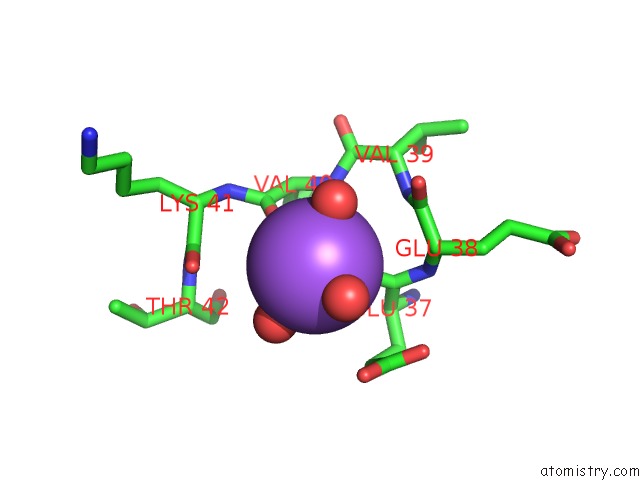
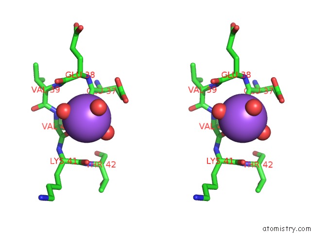
Sodium binding site 1 out of 2 in 4ma5
Go back to
Sodium binding site 1 out
of 2 in the The Crystal Structure of Phosphoribosylaminoimidazole Carboxylase Atpase Subunit of Francisella Tularensis Subsp. Tularensis Schu S4 in Complex with An Atp Analog, Amp-Pnp.
Mono view
Stereo pair view
Mono view
Stereo pair view
A full contact list of Sodium with other atoms in the Na binding
site number 1 of The Crystal Structure of Phosphoribosylaminoimidazole Carboxylase Atpase Subunit of Francisella Tularensis Subsp. Tularensis Schu S4 in Complex with An Atp Analog, Amp-Pnp. within 5.0Å range:
|
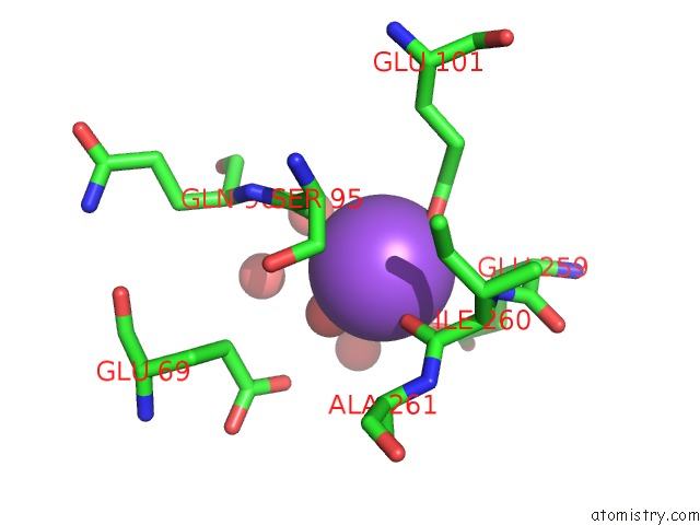
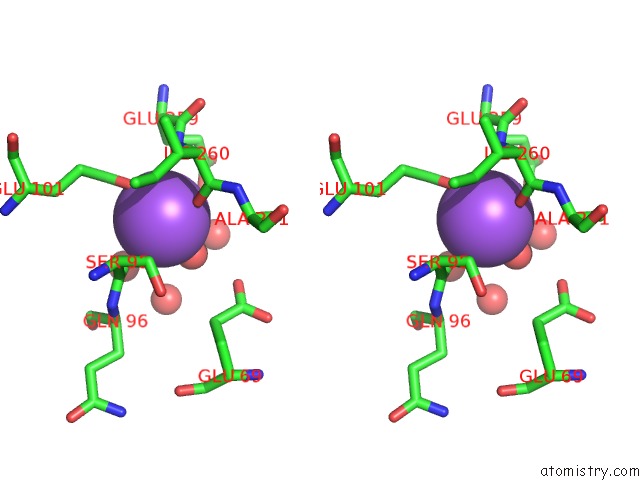
Sodium binding site 2 out of 2 in 4ma5
Go back to
Sodium binding site 2 out
of 2 in the The Crystal Structure of Phosphoribosylaminoimidazole Carboxylase Atpase Subunit of Francisella Tularensis Subsp. Tularensis Schu S4 in Complex with An Atp Analog, Amp-Pnp.
Mono view
Stereo pair view
Mono view
Stereo pair view
A full contact list of Sodium with other atoms in the Na binding
site number 2 of The Crystal Structure of Phosphoribosylaminoimidazole Carboxylase Atpase Subunit of Francisella Tularensis Subsp. Tularensis Schu S4 in Complex with An Atp Analog, Amp-Pnp. within 5.0Å range:
|
Reference:
K.Tan,
M.Zhou,
K.Kwon,
W.F.Anderson,
A.Joachimiak.
The Crystal Structure of Phosphoribosylaminoimidazole Carboxylase Atpase Subunit of Francisella Tularensis Subsp. Tularensis Schu S4 in Complex with An Atp Analog, Amp-Pnp. To Be Published.
Page generated: Mon Oct 7 16:57:56 2024
Last articles
Mg in 4L9ZMg in 4L9Y
Mg in 4LA6
Mg in 4L9W
Mg in 4L81
Mg in 4L9S
Mg in 4L8N
Mg in 4L87
Mg in 4L8G
Mg in 4L80