Sodium »
PDB 8ihy-8jzi »
8iq8 »
Sodium in PDB 8iq8: Crystal Structure of 3,4-Dihydroxyphenylacetate 2,3-Dioxygenase (Dhpao) From Acinetobacter Baumannii
Protein crystallography data
The structure of Crystal Structure of 3,4-Dihydroxyphenylacetate 2,3-Dioxygenase (Dhpao) From Acinetobacter Baumannii, PDB code: 8iq8
was solved by
P.Chitnumsub,
S.Maenpuen,
with X-Ray Crystallography technique. A brief refinement statistics is given in the table below:
| Resolution Low / High (Å) | 21.04 / 1.80 |
| Space group | P 1 21 1 |
| Cell size a, b, c (Å), α, β, γ (°) | 93.433, 62.84, 135.952, 90, 90.09, 90 |
| R / Rfree (%) | 22.5 / 25.5 |
Sodium Binding Sites:
The binding sites of Sodium atom in the Crystal Structure of 3,4-Dihydroxyphenylacetate 2,3-Dioxygenase (Dhpao) From Acinetobacter Baumannii
(pdb code 8iq8). This binding sites where shown within
5.0 Angstroms radius around Sodium atom.
In total 4 binding sites of Sodium where determined in the Crystal Structure of 3,4-Dihydroxyphenylacetate 2,3-Dioxygenase (Dhpao) From Acinetobacter Baumannii, PDB code: 8iq8:
Jump to Sodium binding site number: 1; 2; 3; 4;
In total 4 binding sites of Sodium where determined in the Crystal Structure of 3,4-Dihydroxyphenylacetate 2,3-Dioxygenase (Dhpao) From Acinetobacter Baumannii, PDB code: 8iq8:
Jump to Sodium binding site number: 1; 2; 3; 4;
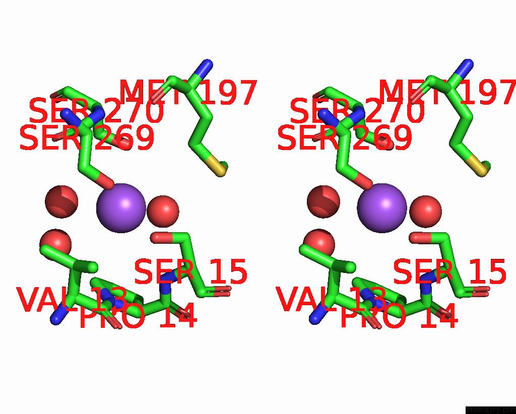
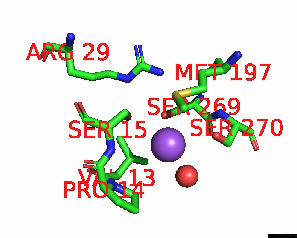
Sodium binding site 1 out of 4 in 8iq8
Go back to
Sodium binding site 1 out
of 4 in the Crystal Structure of 3,4-Dihydroxyphenylacetate 2,3-Dioxygenase (Dhpao) From Acinetobacter Baumannii
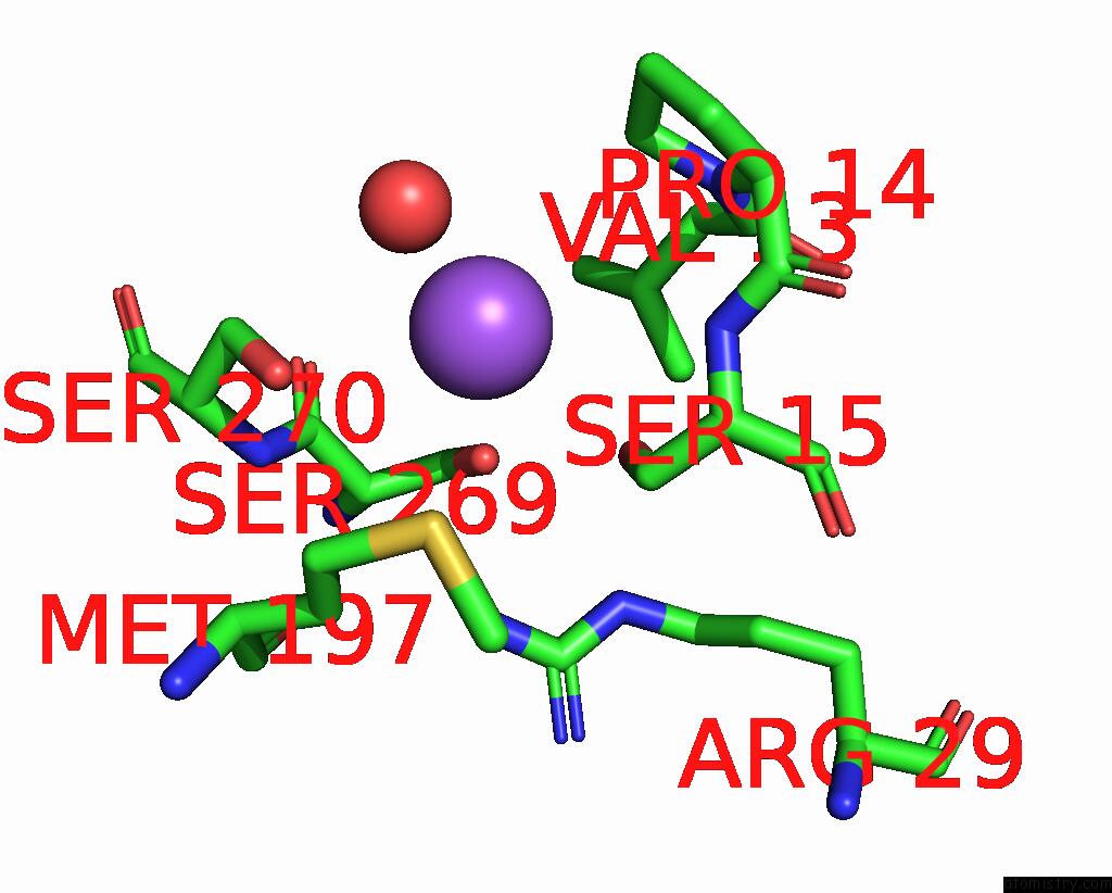
Mono view

Stereo pair view
Mono view
Stereo pair view
A full contact list of Sodium with other atoms in the Na binding
site number 1 of Crystal Structure of 3,4-Dihydroxyphenylacetate 2,3-Dioxygenase (Dhpao) From Acinetobacter Baumannii within 5.0Å range:
|
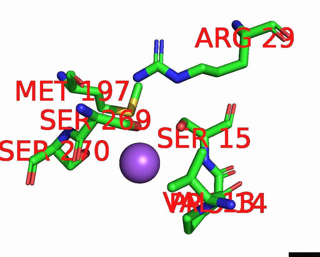
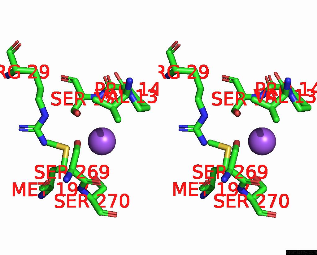
Sodium binding site 2 out of 4 in 8iq8
Go back to
Sodium binding site 2 out
of 4 in the Crystal Structure of 3,4-Dihydroxyphenylacetate 2,3-Dioxygenase (Dhpao) From Acinetobacter Baumannii

Mono view
Stereo pair view
Mono view
Stereo pair view
A full contact list of Sodium with other atoms in the Na binding
site number 2 of Crystal Structure of 3,4-Dihydroxyphenylacetate 2,3-Dioxygenase (Dhpao) From Acinetobacter Baumannii within 5.0Å range:
|
Sodium binding site 3 out of 4 in 8iq8
Go back to
Sodium binding site 3 out
of 4 in the Crystal Structure of 3,4-Dihydroxyphenylacetate 2,3-Dioxygenase (Dhpao) From Acinetobacter Baumannii
Mono view
Stereo pair view
Mono view
Stereo pair view
A full contact list of Sodium with other atoms in the Na binding
site number 3 of Crystal Structure of 3,4-Dihydroxyphenylacetate 2,3-Dioxygenase (Dhpao) From Acinetobacter Baumannii within 5.0Å range:
|
Sodium binding site 4 out of 4 in 8iq8
Go back to
Sodium binding site 4 out
of 4 in the Crystal Structure of 3,4-Dihydroxyphenylacetate 2,3-Dioxygenase (Dhpao) From Acinetobacter Baumannii
Mono view

Stereo pair view
Mono view
Stereo pair view
A full contact list of Sodium with other atoms in the Na binding
site number 4 of Crystal Structure of 3,4-Dihydroxyphenylacetate 2,3-Dioxygenase (Dhpao) From Acinetobacter Baumannii within 5.0Å range:
|
Reference:
P.Pimviriyakul,
S.Buttranon,
S.Soithongcharoen,
C.Supawatkon,
K.Disayabootr,
P.Watthaisong,
R.Tinikul,
A.Jaruwat,
P.Chaiyen,
P.Chitnumsub,
S.Maenpuen.
Structure and Biochemical Characterization of An Extradiol 3,4-Dihydroxyphenylacetate 2,3-Dioxygenase From Acinetobacter Baumannii. Arch.Biochem.Biophys. V. 747 09768 2023.
ISSN: ESSN 1096-0384
PubMed: 37769893
DOI: 10.1016/J.ABB.2023.109768
Page generated: Wed Oct 9 12:34:36 2024
ISSN: ESSN 1096-0384
PubMed: 37769893
DOI: 10.1016/J.ABB.2023.109768
Last articles
Zn in 9JYWZn in 9IR4
Zn in 9IR3
Zn in 9GMX
Zn in 9GMW
Zn in 9JEJ
Zn in 9ERF
Zn in 9ERE
Zn in 9EGV
Zn in 9EGW