Sodium in PDB 7zi3: Crystal Structure of Dck C4S-S74E Mutant in Complex with Udp and the OR0642 Inhibitor
Enzymatic activity of Crystal Structure of Dck C4S-S74E Mutant in Complex with Udp and the OR0642 Inhibitor
All present enzymatic activity of Crystal Structure of Dck C4S-S74E Mutant in Complex with Udp and the OR0642 Inhibitor:
2.7.1.113; 2.7.1.74; 2.7.1.76;
2.7.1.113; 2.7.1.74; 2.7.1.76;
Protein crystallography data
The structure of Crystal Structure of Dck C4S-S74E Mutant in Complex with Udp and the OR0642 Inhibitor, PDB code: 7zi3
was solved by
K.Ben-Yaala,
M.Saez-Ayala,
S.Betzi,
E.Rebuffet,
X.Morelli,
with X-Ray Crystallography technique. A brief refinement statistics is given in the table below:
| Resolution Low / High (Å) | 48.75 / 1.90 |
| Space group | P 41 2 2 |
| Cell size a, b, c (Å), α, β, γ (°) | 68.94, 68.94, 120.763, 90, 90, 90 |
| R / Rfree (%) | 19 / 23 |
Other elements in 7zi3:
The structure of Crystal Structure of Dck C4S-S74E Mutant in Complex with Udp and the OR0642 Inhibitor also contains other interesting chemical elements:
| Fluorine | (F) | 3 atoms |
Sodium Binding Sites:
The binding sites of Sodium atom in the Crystal Structure of Dck C4S-S74E Mutant in Complex with Udp and the OR0642 Inhibitor
(pdb code 7zi3). This binding sites where shown within
5.0 Angstroms radius around Sodium atom.
In total only one binding site of Sodium was determined in the Crystal Structure of Dck C4S-S74E Mutant in Complex with Udp and the OR0642 Inhibitor, PDB code: 7zi3:
In total only one binding site of Sodium was determined in the Crystal Structure of Dck C4S-S74E Mutant in Complex with Udp and the OR0642 Inhibitor, PDB code: 7zi3:
Sodium binding site 1 out of 1 in 7zi3
Go back to
Sodium binding site 1 out
of 1 in the Crystal Structure of Dck C4S-S74E Mutant in Complex with Udp and the OR0642 Inhibitor
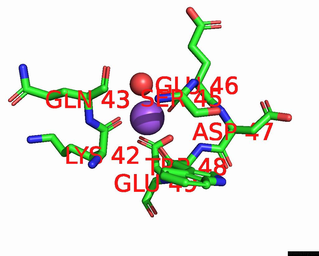
Mono view
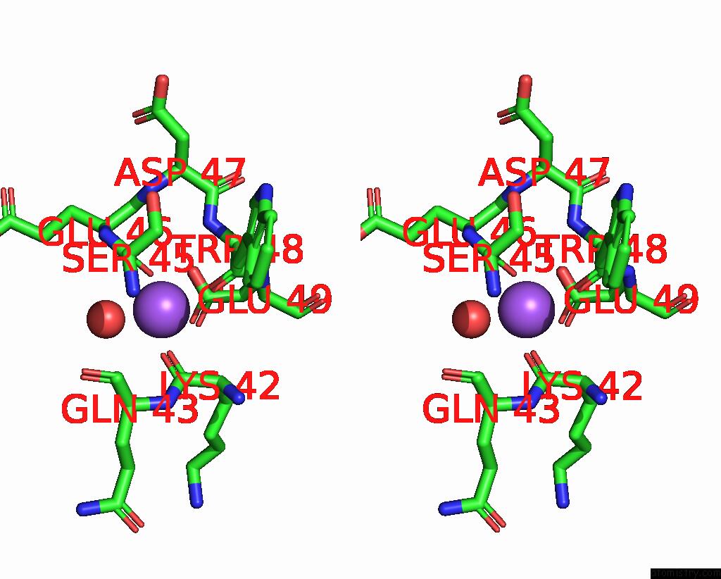
Stereo pair view
Mono view
Stereo pair view
A full contact list of Sodium with other atoms in the Na binding
site number 1 of Crystal Structure of Dck C4S-S74E Mutant in Complex with Udp and the OR0642 Inhibitor within 5.0Å range:
|
Reference:
M.Saez-Ayala,
L.Hoffer,
S.Abel,
K.Ben Yaala,
B.Sicard,
G.P.Andrieu,
M.Latiri,
E.K.Davison,
M.A.Ciufolini,
P.Bremond,
E.Rebuffet,
P.Roche,
C.Derviaux,
E.Voisset,
C.Montersino,
R.Castellano,
Y.Collette,
V.Asnafi,
S.Betzi,
P.Dubreuil,
S.Combes,
X.Morelli.
From A Drug Repositioning to A Structure-Based Drug Design Approach to Tackle Acute Lymphoblastic Leukemia. Nat Commun V. 14 3079 2023.
ISSN: ESSN 2041-1723
PubMed: 37248212
DOI: 10.1038/S41467-023-38668-2
Page generated: Wed Oct 9 10:16:38 2024
ISSN: ESSN 2041-1723
PubMed: 37248212
DOI: 10.1038/S41467-023-38668-2
Last articles
Zn in 9MJ5Zn in 9HNW
Zn in 9G0L
Zn in 9FNE
Zn in 9DZN
Zn in 9E0I
Zn in 9D32
Zn in 9DAK
Zn in 8ZXC
Zn in 8ZUF