Sodium »
PDB 7xft-7ypy »
7ykb »
Sodium in PDB 7ykb: Neutron Structure of Pcya D105N Mutant Complexed with Biliverdin at Room Temperature
Enzymatic activity of Neutron Structure of Pcya D105N Mutant Complexed with Biliverdin at Room Temperature
All present enzymatic activity of Neutron Structure of Pcya D105N Mutant Complexed with Biliverdin at Room Temperature:
1.3.7.5;
1.3.7.5;
Protein crystallography data
The structure of Neutron Structure of Pcya D105N Mutant Complexed with Biliverdin at Room Temperature, PDB code: 7ykb
was solved by
M.Unno,
R.Nanasawa,
with X-Ray Crystallography technique. A brief refinement statistics is given in the table below:
| Resolution Low / High (Å) | N/A / 1.38 |
| Space group | P 21 21 2 |
| Cell size a, b, c (Å), α, β, γ (°) | 71.174, 97.523, 43.277, 90, 90, 90 |
| R / Rfree (%) | 16.6 / 18.1 |
Sodium Binding Sites:
The binding sites of Sodium atom in the Neutron Structure of Pcya D105N Mutant Complexed with Biliverdin at Room Temperature
(pdb code 7ykb). This binding sites where shown within
5.0 Angstroms radius around Sodium atom.
In total only one binding site of Sodium was determined in the Neutron Structure of Pcya D105N Mutant Complexed with Biliverdin at Room Temperature, PDB code: 7ykb:
In total only one binding site of Sodium was determined in the Neutron Structure of Pcya D105N Mutant Complexed with Biliverdin at Room Temperature, PDB code: 7ykb:
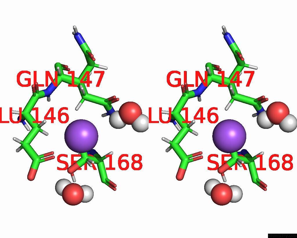
Sodium binding site 1 out of 1 in 7ykb
Go back to
Sodium binding site 1 out
of 1 in the Neutron Structure of Pcya D105N Mutant Complexed with Biliverdin at Room Temperature

Mono view
Stereo pair view
Mono view
Stereo pair view
A full contact list of Sodium with other atoms in the Na binding
site number 1 of Neutron Structure of Pcya D105N Mutant Complexed with Biliverdin at Room Temperature within 5.0Å range:
|
Reference:
T.Joutsuka,
R.Nanasawa,
K.Igarashi,
K.Horie,
M.Sugishima,
Y.Hagiwara,
K.Wada,
K.Fukuyama,
N.Yano,
S.Mori,
A.Ostermann,
K.Kusaka,
M.Unno.
Neutron Crystallography and Quantum Chemical Analysis of Bilin Reductase Pcya Mutants Reveal Substrate and Catalytic Residue Protonation States. J.Biol.Chem. V. 299 02763 2022.
ISSN: ESSN 1083-351X
PubMed: 36463961
DOI: 10.1016/J.JBC.2022.102763
Page generated: Wed Oct 9 09:54:52 2024
ISSN: ESSN 1083-351X
PubMed: 36463961
DOI: 10.1016/J.JBC.2022.102763
Last articles
Zn in 9JYWZn in 9IR4
Zn in 9IR3
Zn in 9GMX
Zn in 9GMW
Zn in 9JEJ
Zn in 9ERF
Zn in 9ERE
Zn in 9EGV
Zn in 9EGW