Sodium »
PDB 7xft-7ypy »
7xsf »
Sodium in PDB 7xsf: Crystal Structure of CLAGL29A
Protein crystallography data
The structure of Crystal Structure of CLAGL29A, PDB code: 7xsf
was solved by
R.Shishiuchi,
H.Kang,
T.Tagami,
M.Okuyama,
with X-Ray Crystallography technique. A brief refinement statistics is given in the table below:
| Resolution Low / High (Å) | 47.81 / 2.01 |
| Space group | P 1 21 1 |
| Cell size a, b, c (Å), α, β, γ (°) | 74.577, 95.521, 83.55, 90, 97.33, 90 |
| R / Rfree (%) | 20.5 / 24.3 |
Sodium Binding Sites:
The binding sites of Sodium atom in the Crystal Structure of CLAGL29A
(pdb code 7xsf). This binding sites where shown within
5.0 Angstroms radius around Sodium atom.
In total 2 binding sites of Sodium where determined in the Crystal Structure of CLAGL29A, PDB code: 7xsf:
Jump to Sodium binding site number: 1; 2;
In total 2 binding sites of Sodium where determined in the Crystal Structure of CLAGL29A, PDB code: 7xsf:
Jump to Sodium binding site number: 1; 2;
Sodium binding site 1 out of 2 in 7xsf
Go back to
Sodium binding site 1 out
of 2 in the Crystal Structure of CLAGL29A
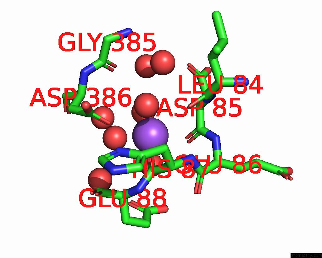
Mono view

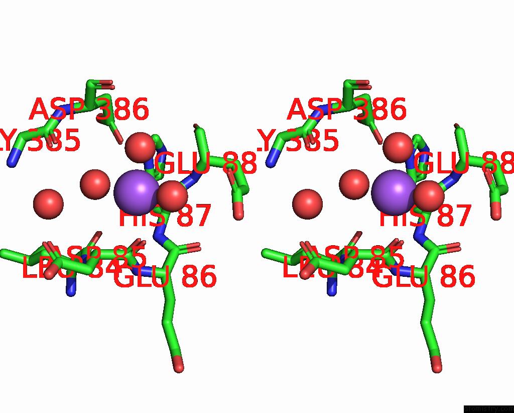
Stereo pair view
Mono view
Stereo pair view
A full contact list of Sodium with other atoms in the Na binding
site number 1 of Crystal Structure of CLAGL29A within 5.0Å range:
|
Sodium binding site 2 out of 2 in 7xsf
Go back to
Sodium binding site 2 out
of 2 in the Crystal Structure of CLAGL29A
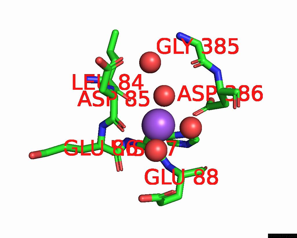
Mono view
Stereo pair view
Mono view
Stereo pair view
A full contact list of Sodium with other atoms in the Na binding
site number 2 of Crystal Structure of CLAGL29A within 5.0Å range:
|
Reference:
R.Shishiuchi,
H.Kang,
T.Tagami,
Y.Ueda,
W.Lang,
A.Kimura,
M.Okuyama.
Discovery of Alpha-L-Glucosidase Raises the Possibility of Alpha-L-Glucosides in Nature. Acs Omega V. 7 47411 2022.
ISSN: ESSN 2470-1343
PubMed: 36570207
DOI: 10.1021/ACSOMEGA.2C06991
Page generated: Wed Oct 9 09:49:58 2024
ISSN: ESSN 2470-1343
PubMed: 36570207
DOI: 10.1021/ACSOMEGA.2C06991
Last articles
Zn in 9JYWZn in 9IR4
Zn in 9IR3
Zn in 9GMX
Zn in 9GMW
Zn in 9JEJ
Zn in 9ERF
Zn in 9ERE
Zn in 9EGV
Zn in 9EGW