Sodium »
PDB 7m6k-7mq3 »
7mfp »
Sodium in PDB 7mfp: Crystal Structure of the L136 Aminotransferase K185A From Acanthamoeba Polyphaga Mimivirus in the Presence of the Udp-Viosamine External Aldimine
Protein crystallography data
The structure of Crystal Structure of the L136 Aminotransferase K185A From Acanthamoeba Polyphaga Mimivirus in the Presence of the Udp-Viosamine External Aldimine, PDB code: 7mfp
was solved by
C.A.Seltzner,
J.B.Thoden,
H.M.Holden,
with X-Ray Crystallography technique. A brief refinement statistics is given in the table below:
| Resolution Low / High (Å) | 29.72 / 1.85 |
| Space group | P 21 21 21 |
| Cell size a, b, c (Å), α, β, γ (°) | 91.033, 108.548, 145.514, 90, 90, 90 |
| R / Rfree (%) | 17.2 / 21.9 |
Other elements in 7mfp:
The structure of Crystal Structure of the L136 Aminotransferase K185A From Acanthamoeba Polyphaga Mimivirus in the Presence of the Udp-Viosamine External Aldimine also contains other interesting chemical elements:
| Chlorine | (Cl) | 8 atoms |
Sodium Binding Sites:
The binding sites of Sodium atom in the Crystal Structure of the L136 Aminotransferase K185A From Acanthamoeba Polyphaga Mimivirus in the Presence of the Udp-Viosamine External Aldimine
(pdb code 7mfp). This binding sites where shown within
5.0 Angstroms radius around Sodium atom.
In total 2 binding sites of Sodium where determined in the Crystal Structure of the L136 Aminotransferase K185A From Acanthamoeba Polyphaga Mimivirus in the Presence of the Udp-Viosamine External Aldimine, PDB code: 7mfp:
Jump to Sodium binding site number: 1; 2;
In total 2 binding sites of Sodium where determined in the Crystal Structure of the L136 Aminotransferase K185A From Acanthamoeba Polyphaga Mimivirus in the Presence of the Udp-Viosamine External Aldimine, PDB code: 7mfp:
Jump to Sodium binding site number: 1; 2;
Sodium binding site 1 out of 2 in 7mfp
Go back to
Sodium binding site 1 out
of 2 in the Crystal Structure of the L136 Aminotransferase K185A From Acanthamoeba Polyphaga Mimivirus in the Presence of the Udp-Viosamine External Aldimine

Mono view
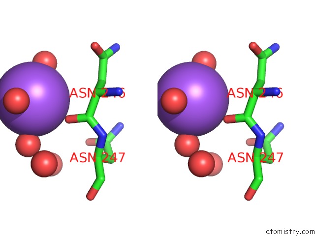
Stereo pair view
Mono view
Stereo pair view
A full contact list of Sodium with other atoms in the Na binding
site number 1 of Crystal Structure of the L136 Aminotransferase K185A From Acanthamoeba Polyphaga Mimivirus in the Presence of the Udp-Viosamine External Aldimine within 5.0Å range:
|
Sodium binding site 2 out of 2 in 7mfp
Go back to
Sodium binding site 2 out
of 2 in the Crystal Structure of the L136 Aminotransferase K185A From Acanthamoeba Polyphaga Mimivirus in the Presence of the Udp-Viosamine External Aldimine

Mono view

Stereo pair view
Mono view
Stereo pair view
A full contact list of Sodium with other atoms in the Na binding
site number 2 of Crystal Structure of the L136 Aminotransferase K185A From Acanthamoeba Polyphaga Mimivirus in the Presence of the Udp-Viosamine External Aldimine within 5.0Å range:
|
Reference:
C.A.Seltzner,
J.D.Ferek,
J.B.Thoden,
H.M.Holden.
Characterization of An Aminotransferase From Acanthamoeba Polyphaga Mimivirus To Be Published.
Page generated: Tue Oct 8 17:57:43 2024
Last articles
Zn in 9JYWZn in 9IR4
Zn in 9IR3
Zn in 9GMX
Zn in 9GMW
Zn in 9JEJ
Zn in 9ERF
Zn in 9ERE
Zn in 9EGV
Zn in 9EGW