Sodium in PDB 7djc: Crystal Structure of the G26C/Q250A Mutant of Leut
Protein crystallography data
The structure of Crystal Structure of the G26C/Q250A Mutant of Leut, PDB code: 7djc
was solved by
J.Fan,
Y.Xiao,
Z.Sun,
X.Zhou,
with X-Ray Crystallography technique. A brief refinement statistics is given in the table below:
Resolution Low / High (Å)
|
31.51 /
2.70
|
Space group
|
C 1 2 1
|
Cell size a, b, c (Å), α, β, γ (°)
|
90.758,
87.606,
81.813,
90,
94.35,
90
|
R / Rfree (%)
|
20.6 /
24.2
|
Sodium Binding Sites:
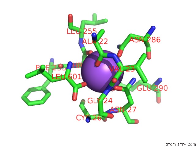
The binding sites of Sodium atom in the Crystal Structure of the G26C/Q250A Mutant of Leut
(pdb code 7djc). This binding sites where shown within
5.0 Angstroms radius around Sodium atom.
In total 2 binding sites of Sodium where determined in the
Crystal Structure of the G26C/Q250A Mutant of Leut, PDB code: 7djc:
Jump to Sodium binding site number:
1;
2;
Sodium binding site 1 out
of 2 in 7djc
Go back to
Sodium Binding Sites List in 7djc
Sodium binding site 1 out
of 2 in the Crystal Structure of the G26C/Q250A Mutant of Leut
 Mono view
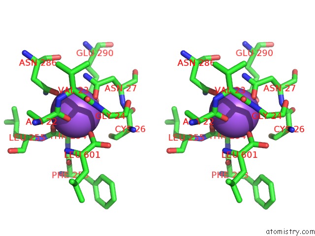
 Stereo pair view
|
A full contact list of Sodium with other atoms in the Na binding
site number 1 of Crystal Structure of the G26C/Q250A Mutant of Leut within 5.0Å range:
probe
|
atom
|
residue
|
distance (Å)
|
B
|
Occ
|
A:Na602
b:74.2
occ:1.00
|
OD1
|
A:ASN27
|
2.4
|
60.9
|
1.0
|
O
|
A:THR254
|
2.4
|
55.2
|
1.0
|
OG1
|
A:THR254
|
2.4
|
55.6
|
1.0
|
O
|
A:ALA22
|
2.4
|
54.1
|
1.0
|
OXT
|
A:LEU601
|
2.4
|
55.0
|
1.0
|
OD1
|
A:ASN286
|
2.5
|
46.2
|
1.0
|
C
|
A:THR254
|
3.1
|
55.3
|
1.0
|
CA
|
A:THR254
|
3.1
|
54.5
|
1.0
|
CG
|
A:ASN27
|
3.2
|
57.2
|
1.0
|
CB
|
A:THR254
|
3.3
|
56.5
|
1.0
|
C
|
A:ALA22
|
3.3
|
52.0
|
1.0
|
CG
|
A:ASN286
|
3.4
|
45.0
|
1.0
|
ND2
|
A:ASN27
|
3.4
|
47.1
|
1.0
|
C
|
A:LEU601
|
3.6
|
54.0
|
1.0
|
ND2
|
A:ASN286
|
3.7
|
42.2
|
1.0
|
N
|
A:LEU601
|
3.8
|
51.5
|
1.0
|
N
|
A:GLY24
|
3.9
|
47.0
|
1.0
|
CA
|
A:VAL23
|
4.0
|
53.3
|
1.0
|
OE2
|
A:GLU290
|
4.0
|
71.7
|
1.0
|
N
|
A:VAL23
|
4.0
|
55.2
|
1.0
|
CA
|
A:LEU601
|
4.3
|
52.7
|
1.0
|
CA
|
A:ALA22
|
4.3
|
49.7
|
1.0
|
N
|
A:LEU255
|
4.4
|
48.3
|
1.0
|
N
|
A:ASN27
|
4.4
|
50.1
|
1.0
|
C
|
A:VAL23
|
4.4
|
54.0
|
1.0
|
O
|
A:LEU601
|
4.5
|
54.6
|
1.0
|
CG2
|
A:THR254
|
4.5
|
56.5
|
1.0
|
N
|
A:THR254
|
4.5
|
52.3
|
1.0
|
CB
|
A:ASN27
|
4.5
|
57.4
|
1.0
|
CB
|
A:CYS26
|
4.5
|
49.2
|
1.0
|
CB
|
A:ALA22
|
4.6
|
41.4
|
1.0
|
O
|
A:PHE253
|
4.7
|
53.0
|
1.0
|
CD
|
A:GLU290
|
4.8
|
71.1
|
1.0
|
CA
|
A:ASN27
|
4.8
|
57.3
|
1.0
|
CB
|
A:ASN286
|
4.8
|
50.5
|
1.0
|
CA
|
A:GLY24
|
4.9
|
43.3
|
1.0
|
SG
|
A:CYS26
|
4.9
|
54.4
|
1.0
|
|
Sodium binding site 2 out
of 2 in 7djc
Go back to
Sodium Binding Sites List in 7djc
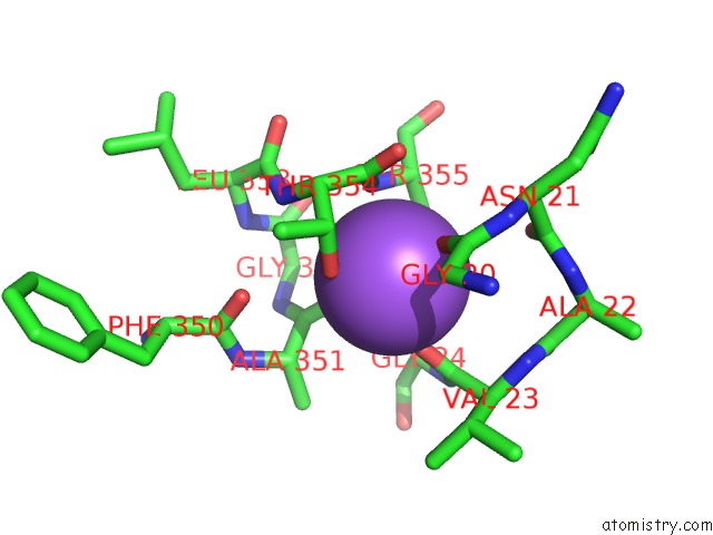
Sodium binding site 2 out
of 2 in the Crystal Structure of the G26C/Q250A Mutant of Leut
 Mono view
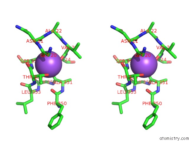
 Stereo pair view
|
A full contact list of Sodium with other atoms in the Na binding
site number 2 of Crystal Structure of the G26C/Q250A Mutant of Leut within 5.0Å range:
probe
|
atom
|
residue
|
distance (Å)
|
B
|
Occ
|
A:Na603
b:50.1
occ:1.00
|
O
|
A:VAL23
|
2.3
|
57.9
|
1.0
|
O
|
A:ALA351
|
2.3
|
55.7
|
1.0
|
O
|
A:GLY20
|
2.3
|
54.4
|
1.0
|
OG1
|
A:THR354
|
2.3
|
34.5
|
1.0
|
OG
|
A:SER355
|
2.4
|
53.3
|
1.0
|
N
|
A:SER355
|
2.8
|
47.9
|
1.0
|
CB
|
A:SER355
|
3.1
|
47.8
|
1.0
|
C
|
A:THR354
|
3.2
|
45.1
|
1.0
|
CB
|
A:THR354
|
3.3
|
41.7
|
1.0
|
C
|
A:VAL23
|
3.3
|
54.0
|
1.0
|
CA
|
A:SER355
|
3.4
|
47.1
|
1.0
|
C
|
A:ALA351
|
3.4
|
53.7
|
1.0
|
C
|
A:GLY20
|
3.5
|
53.2
|
1.0
|
CA
|
A:THR354
|
3.6
|
45.0
|
1.0
|
CA
|
A:GLY24
|
3.7
|
43.3
|
1.0
|
O
|
A:THR354
|
3.8
|
43.3
|
1.0
|
N
|
A:THR354
|
3.9
|
49.8
|
1.0
|
N
|
A:GLY24
|
3.9
|
47.0
|
1.0
|
CA
|
A:ALA351
|
4.0
|
46.3
|
1.0
|
CA
|
A:GLY20
|
4.1
|
50.0
|
1.0
|
O
|
A:ASN21
|
4.4
|
48.4
|
1.0
|
O
|
A:PHE350
|
4.4
|
51.8
|
1.0
|
N
|
A:GLY352
|
4.5
|
51.5
|
1.0
|
CA
|
A:VAL23
|
4.5
|
53.3
|
1.0
|
N
|
A:ASN21
|
4.5
|
53.9
|
1.0
|
N
|
A:VAL23
|
4.6
|
55.2
|
1.0
|
C
|
A:ASN21
|
4.6
|
47.6
|
1.0
|
C
|
A:GLY352
|
4.6
|
46.9
|
1.0
|
O
|
A:GLY352
|
4.6
|
50.9
|
1.0
|
CG2
|
A:THR354
|
4.7
|
41.4
|
1.0
|
CA
|
A:GLY352
|
4.7
|
48.5
|
1.0
|
CA
|
A:ASN21
|
4.8
|
49.9
|
1.0
|
C
|
A:SER355
|
4.9
|
53.2
|
1.0
|
CB
|
A:ALA351
|
4.9
|
40.7
|
1.0
|
C
|
A:LEU353
|
4.9
|
55.6
|
1.0
|
C
|
A:ALA22
|
4.9
|
52.0
|
1.0
|
CB
|
A:VAL23
|
5.0
|
55.0
|
1.0
|
|
Reference:
J.Fan,
Y.Xiao,
M.Quick,
Y.Yang,
Z.Sun,
J.A.Javitch,
X.Zhou.
Crystal Structures of Leut Reveal Conformational Dynamics in the Outward-Facing States J.Biol.Chem. 2021.
ISSN: ESSN 1083-351X
Page generated: Tue Oct 8 16:35:17 2024
|