Sodium »
PDB 7cfs-7d5w »
7cnw »
Sodium in PDB 7cnw: Crystal Structure of Apo Psd From E. Coli (1.90 A)
Enzymatic activity of Crystal Structure of Apo Psd From E. Coli (1.90 A)
All present enzymatic activity of Crystal Structure of Apo Psd From E. Coli (1.90 A):
4.1.1.65;
4.1.1.65;
Protein crystallography data
The structure of Crystal Structure of Apo Psd From E. Coli (1.90 A), PDB code: 7cnw
was solved by
J.Kim,
G.Cho,
with X-Ray Crystallography technique. A brief refinement statistics is given in the table below:
| Resolution Low / High (Å) | 41.81 / 1.90 |
| Space group | P 21 21 21 |
| Cell size a, b, c (Å), α, β, γ (°) | 77.443, 79.817, 147.089, 90, 90, 90 |
| R / Rfree (%) | 21.2 / 24.3 |
Sodium Binding Sites:
The binding sites of Sodium atom in the Crystal Structure of Apo Psd From E. Coli (1.90 A)
(pdb code 7cnw). This binding sites where shown within
5.0 Angstroms radius around Sodium atom.
In total 2 binding sites of Sodium where determined in the Crystal Structure of Apo Psd From E. Coli (1.90 A), PDB code: 7cnw:
Jump to Sodium binding site number: 1; 2;
In total 2 binding sites of Sodium where determined in the Crystal Structure of Apo Psd From E. Coli (1.90 A), PDB code: 7cnw:
Jump to Sodium binding site number: 1; 2;
Sodium binding site 1 out of 2 in 7cnw
Go back to
Sodium binding site 1 out
of 2 in the Crystal Structure of Apo Psd From E. Coli (1.90 A)
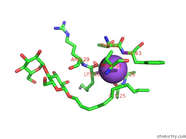
Mono view
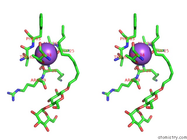
Stereo pair view
Mono view
Stereo pair view
A full contact list of Sodium with other atoms in the Na binding
site number 1 of Crystal Structure of Apo Psd From E. Coli (1.90 A) within 5.0Å range:
|
Sodium binding site 2 out of 2 in 7cnw
Go back to
Sodium binding site 2 out
of 2 in the Crystal Structure of Apo Psd From E. Coli (1.90 A)
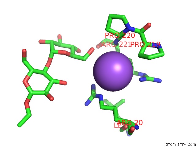
Mono view
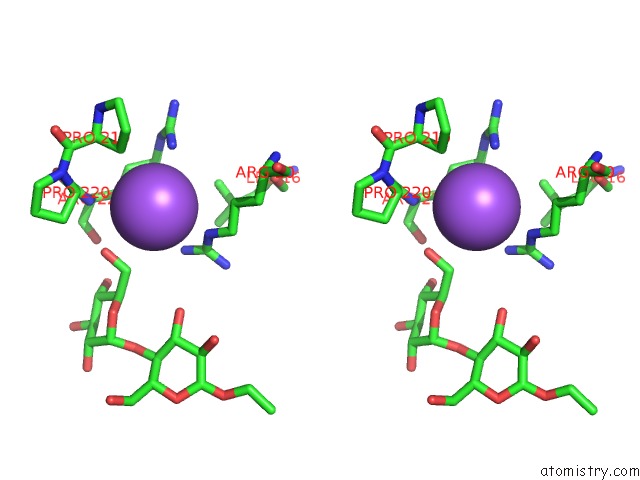
Stereo pair view
Mono view
Stereo pair view
A full contact list of Sodium with other atoms in the Na binding
site number 2 of Crystal Structure of Apo Psd From E. Coli (1.90 A) within 5.0Å range:
|
Reference:
G.Cho,
E.Lee,
J.Kim.
Structural Insights Into Phosphatidylethanolamine Formation in Bacterial Membrane Biogenesis. Sci Rep V. 11 5785 2021.
ISSN: ESSN 2045-2322
PubMed: 33707636
DOI: 10.1038/S41598-021-85195-5
Page generated: Tue Oct 8 16:23:56 2024
ISSN: ESSN 2045-2322
PubMed: 33707636
DOI: 10.1038/S41598-021-85195-5
Last articles
Cl in 5IA3Cl in 5IAE
Cl in 5IBN
Cl in 5IBC
Cl in 5IB5
Cl in 5IAB
Cl in 5IA0
Cl in 5I7A
Cl in 5IA1
Cl in 5I9Y