Sodium »
PDB 6ycs-6yt4 »
6ymk »
Sodium in PDB 6ymk: Crystal Structure of the Sam-Sah Riboswitch with Amp
Protein crystallography data
The structure of Crystal Structure of the Sam-Sah Riboswitch with Amp, PDB code: 6ymk
was solved by
L.Huang,
D.M.J.Lilley,
with X-Ray Crystallography technique. A brief refinement statistics is given in the table below:
| Resolution Low / High (Å) | 37.48 / 2.03 |
| Space group | C 1 2 1 |
| Cell size a, b, c (Å), α, β, γ (°) | 87.100, 147.847, 75.009, 90.00, 91.84, 90.00 |
| R / Rfree (%) | 22.5 / 26.4 |
Other elements in 6ymk:
The structure of Crystal Structure of the Sam-Sah Riboswitch with Amp also contains other interesting chemical elements:
| Bromine | (Br) | 6 atoms |
Sodium Binding Sites:
The binding sites of Sodium atom in the Crystal Structure of the Sam-Sah Riboswitch with Amp
(pdb code 6ymk). This binding sites where shown within
5.0 Angstroms radius around Sodium atom.
In total 2 binding sites of Sodium where determined in the Crystal Structure of the Sam-Sah Riboswitch with Amp, PDB code: 6ymk:
Jump to Sodium binding site number: 1; 2;
In total 2 binding sites of Sodium where determined in the Crystal Structure of the Sam-Sah Riboswitch with Amp, PDB code: 6ymk:
Jump to Sodium binding site number: 1; 2;
Sodium binding site 1 out of 2 in 6ymk
Go back to
Sodium binding site 1 out
of 2 in the Crystal Structure of the Sam-Sah Riboswitch with Amp
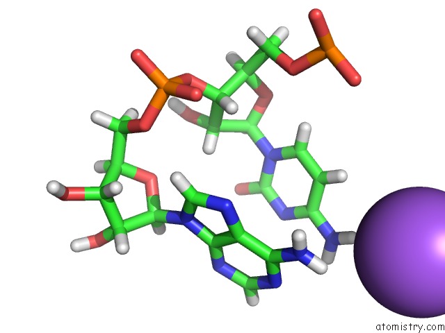
Mono view
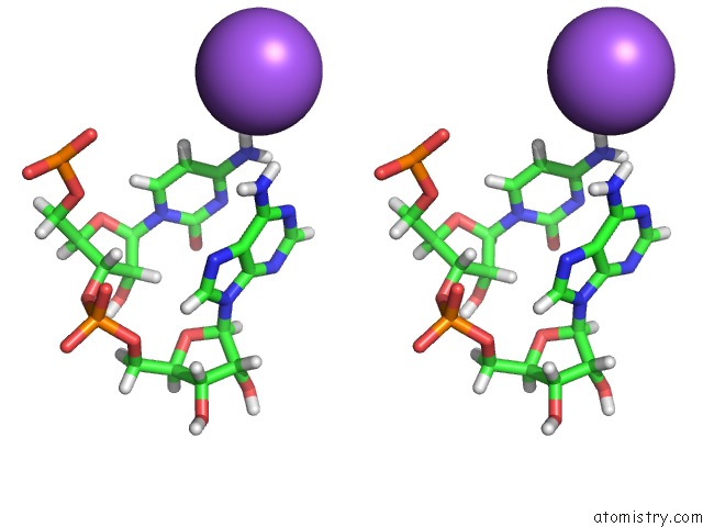
Stereo pair view
Mono view
Stereo pair view
A full contact list of Sodium with other atoms in the Na binding
site number 1 of Crystal Structure of the Sam-Sah Riboswitch with Amp within 5.0Å range:
|
Sodium binding site 2 out of 2 in 6ymk
Go back to
Sodium binding site 2 out
of 2 in the Crystal Structure of the Sam-Sah Riboswitch with Amp
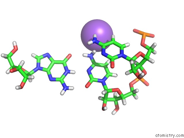
Mono view
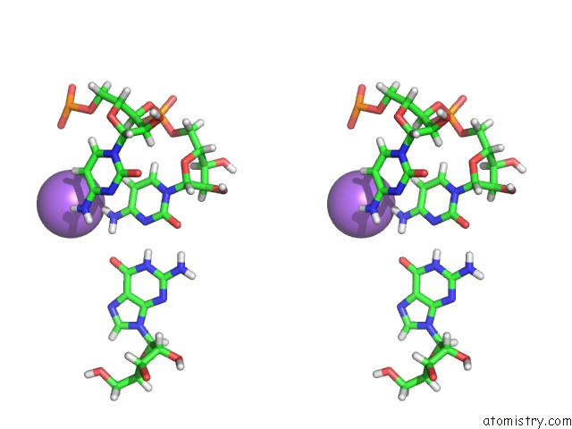
Stereo pair view
Mono view
Stereo pair view
A full contact list of Sodium with other atoms in the Na binding
site number 2 of Crystal Structure of the Sam-Sah Riboswitch with Amp within 5.0Å range:
|
Reference:
L.Huang,
T.W.Liao,
J.Wang,
T.Ha,
D.M.J.Lilley.
Crystal Structure and Ligand-Induced Folding of the Sam/Sah Riboswitch. Nucleic Acids Res. V. 48 7545 2020.
ISSN: ESSN 1362-4962
PubMed: 32520325
DOI: 10.1093/NAR/GKAA493
Page generated: Tue Oct 8 15:10:09 2024
ISSN: ESSN 1362-4962
PubMed: 32520325
DOI: 10.1093/NAR/GKAA493
Last articles
Cl in 5G69Cl in 5G6C
Cl in 5G6A
Cl in 5G6B
Cl in 5G67
Cl in 5G68
Cl in 5G66
Cl in 5G65
Cl in 5G5N
Cl in 5G5O