Sodium »
PDB 6w0s-6wn2 »
6w3l »
Sodium in PDB 6w3l: APE1 Exonuclease Substrate Complex Wild-Type
Enzymatic activity of APE1 Exonuclease Substrate Complex Wild-Type
All present enzymatic activity of APE1 Exonuclease Substrate Complex Wild-Type:
4.2.99.18;
4.2.99.18;
Protein crystallography data
The structure of APE1 Exonuclease Substrate Complex Wild-Type, PDB code: 6w3l
was solved by
B.D.Freudenthal,
A.M.Whitaker,
with X-Ray Crystallography technique. A brief refinement statistics is given in the table below:
| Resolution Low / High (Å) | 24.77 / 2.59 |
| Space group | P 1 21 1 |
| Cell size a, b, c (Å), α, β, γ (°) | 71.371, 65.196, 91.258, 90.00, 110.27, 90.00 |
| R / Rfree (%) | 21.7 / 27.7 |
Other elements in 6w3l:
The structure of APE1 Exonuclease Substrate Complex Wild-Type also contains other interesting chemical elements:
| Calcium | (Ca) | 1 atom |
Sodium Binding Sites:
The binding sites of Sodium atom in the APE1 Exonuclease Substrate Complex Wild-Type
(pdb code 6w3l). This binding sites where shown within
5.0 Angstroms radius around Sodium atom.
In total 2 binding sites of Sodium where determined in the APE1 Exonuclease Substrate Complex Wild-Type, PDB code: 6w3l:
Jump to Sodium binding site number: 1; 2;
In total 2 binding sites of Sodium where determined in the APE1 Exonuclease Substrate Complex Wild-Type, PDB code: 6w3l:
Jump to Sodium binding site number: 1; 2;
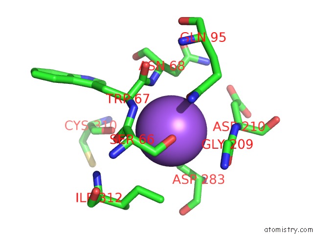
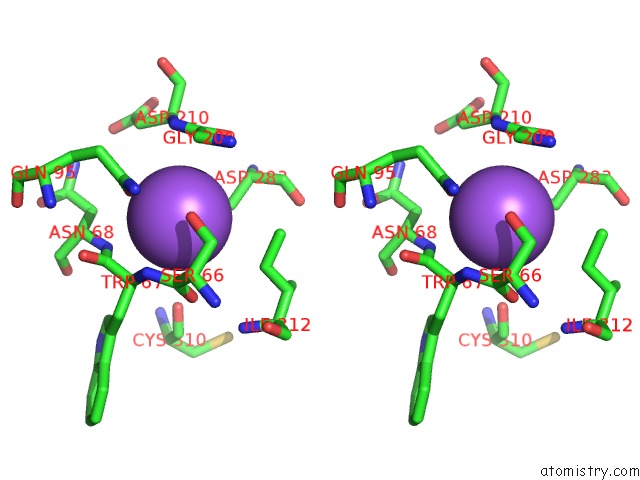
Sodium binding site 1 out of 2 in 6w3l
Go back to
Sodium binding site 1 out
of 2 in the APE1 Exonuclease Substrate Complex Wild-Type

Mono view

Stereo pair view
Mono view
Stereo pair view
A full contact list of Sodium with other atoms in the Na binding
site number 1 of APE1 Exonuclease Substrate Complex Wild-Type within 5.0Å range:
|
Sodium binding site 2 out of 2 in 6w3l
Go back to
Sodium binding site 2 out
of 2 in the APE1 Exonuclease Substrate Complex Wild-Type
Mono view
Stereo pair view
Mono view
Stereo pair view
A full contact list of Sodium with other atoms in the Na binding
site number 2 of APE1 Exonuclease Substrate Complex Wild-Type within 5.0Å range:
|
Reference:
A.M.Whitaker,
W.J.Stark,
T.S.Flynn,
B.D.Freudenthal.
Molecular and Structural Characterization of Disease-Associated APE1 Polymorphisms. Dna Repair (Amst.) V.1-92 02867 2020.
ISSN: ISSN 1568-7856
PubMed: 32454397
DOI: 10.1016/J.DNAREP.2020.102867
Page generated: Tue Oct 8 14:31:24 2024
ISSN: ISSN 1568-7856
PubMed: 32454397
DOI: 10.1016/J.DNAREP.2020.102867
Last articles
Zn in 9J0NZn in 9J0O
Zn in 9J0P
Zn in 9FJX
Zn in 9EKB
Zn in 9C0F
Zn in 9CAH
Zn in 9CH0
Zn in 9CH3
Zn in 9CH1