Sodium »
PDB 6vf7-6w0r »
6vjp »
Sodium in PDB 6vjp: Structure of Staphylococcus Aureus Peptidoglycan O-Acetyltransferase A (Oata) C-Terminal Catalytic Domain
Protein crystallography data
The structure of Structure of Staphylococcus Aureus Peptidoglycan O-Acetyltransferase A (Oata) C-Terminal Catalytic Domain, PDB code: 6vjp
was solved by
C.J.Jones,
D.Sychantha,
P.L.Howell,
A.J.Clarke,
with X-Ray Crystallography technique. A brief refinement statistics is given in the table below:
| Resolution Low / High (Å) | 28.78 / 1.71 |
| Space group | P 1 21 1 |
| Cell size a, b, c (Å), α, β, γ (°) | 42.347, 61.303, 67.690, 90.00, 100.90, 90.00 |
| R / Rfree (%) | 16.6 / 19.5 |
Sodium Binding Sites:
The binding sites of Sodium atom in the Structure of Staphylococcus Aureus Peptidoglycan O-Acetyltransferase A (Oata) C-Terminal Catalytic Domain
(pdb code 6vjp). This binding sites where shown within
5.0 Angstroms radius around Sodium atom.
In total 2 binding sites of Sodium where determined in the Structure of Staphylococcus Aureus Peptidoglycan O-Acetyltransferase A (Oata) C-Terminal Catalytic Domain, PDB code: 6vjp:
Jump to Sodium binding site number: 1; 2;
In total 2 binding sites of Sodium where determined in the Structure of Staphylococcus Aureus Peptidoglycan O-Acetyltransferase A (Oata) C-Terminal Catalytic Domain, PDB code: 6vjp:
Jump to Sodium binding site number: 1; 2;
Sodium binding site 1 out of 2 in 6vjp
Go back to
Sodium binding site 1 out
of 2 in the Structure of Staphylococcus Aureus Peptidoglycan O-Acetyltransferase A (Oata) C-Terminal Catalytic Domain
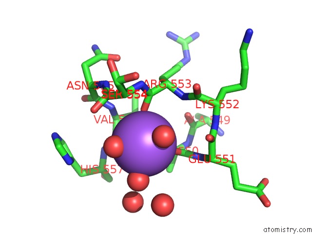
Mono view
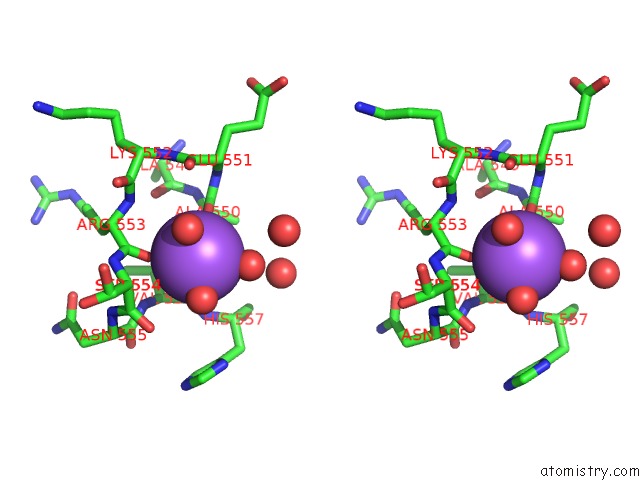
Stereo pair view
Mono view
Stereo pair view
A full contact list of Sodium with other atoms in the Na binding
site number 1 of Structure of Staphylococcus Aureus Peptidoglycan O-Acetyltransferase A (Oata) C-Terminal Catalytic Domain within 5.0Å range:
|
Sodium binding site 2 out of 2 in 6vjp
Go back to
Sodium binding site 2 out
of 2 in the Structure of Staphylococcus Aureus Peptidoglycan O-Acetyltransferase A (Oata) C-Terminal Catalytic Domain
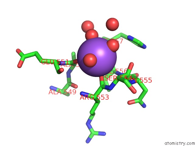
Mono view
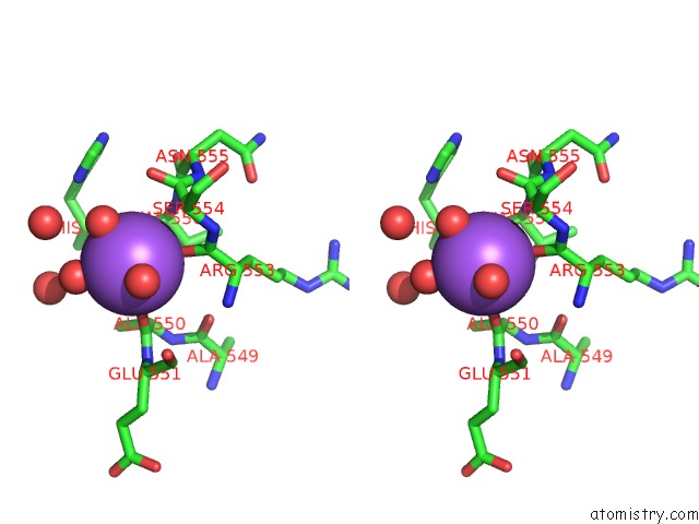
Stereo pair view
Mono view
Stereo pair view
A full contact list of Sodium with other atoms in the Na binding
site number 2 of Structure of Staphylococcus Aureus Peptidoglycan O-Acetyltransferase A (Oata) C-Terminal Catalytic Domain within 5.0Å range:
|
Reference:
C.S.Jones,
D.Sychantha,
P.L.Howell,
A.J.Clarke.
Structural Basis For the O-Acetyltransferase Function of the Extracytoplasmic Domain of Oata From Staphylococcus Aureus J.Biol.Chem. 2020.
ISSN: ESSN 1083-351X
Page generated: Tue Oct 8 14:23:35 2024
ISSN: ESSN 1083-351X
Last articles
Zn in 9J0NZn in 9J0O
Zn in 9J0P
Zn in 9FJX
Zn in 9EKB
Zn in 9C0F
Zn in 9CAH
Zn in 9CH0
Zn in 9CH3
Zn in 9CH1