Sodium »
PDB 6u6u-6v04 »
6uol »
Sodium in PDB 6uol: Y271G Dna Polymerase Beta Substrate Complex with A Templating Cytosine and Incoming Rgtp
Enzymatic activity of Y271G Dna Polymerase Beta Substrate Complex with A Templating Cytosine and Incoming Rgtp
All present enzymatic activity of Y271G Dna Polymerase Beta Substrate Complex with A Templating Cytosine and Incoming Rgtp:
2.7.7.7;
2.7.7.7;
Protein crystallography data
The structure of Y271G Dna Polymerase Beta Substrate Complex with A Templating Cytosine and Incoming Rgtp, PDB code: 6uol
was solved by
M.R.Smith,
B.D.Freudenthal,
with X-Ray Crystallography technique. A brief refinement statistics is given in the table below:
| Resolution Low / High (Å) | 25.09 / 1.94 |
| Space group | P 1 21 1 |
| Cell size a, b, c (Å), α, β, γ (°) | 50.800, 80.400, 55.500, 90.00, 107.90, 90.00 |
| R / Rfree (%) | 18.7 / 25 |
Other elements in 6uol:
The structure of Y271G Dna Polymerase Beta Substrate Complex with A Templating Cytosine and Incoming Rgtp also contains other interesting chemical elements:
| Manganese | (Mn) | 5 atoms |
| Chlorine | (Cl) | 1 atom |
Sodium Binding Sites:
The binding sites of Sodium atom in the Y271G Dna Polymerase Beta Substrate Complex with A Templating Cytosine and Incoming Rgtp
(pdb code 6uol). This binding sites where shown within
5.0 Angstroms radius around Sodium atom.
In total 3 binding sites of Sodium where determined in the Y271G Dna Polymerase Beta Substrate Complex with A Templating Cytosine and Incoming Rgtp, PDB code: 6uol:
Jump to Sodium binding site number: 1; 2; 3;
In total 3 binding sites of Sodium where determined in the Y271G Dna Polymerase Beta Substrate Complex with A Templating Cytosine and Incoming Rgtp, PDB code: 6uol:
Jump to Sodium binding site number: 1; 2; 3;
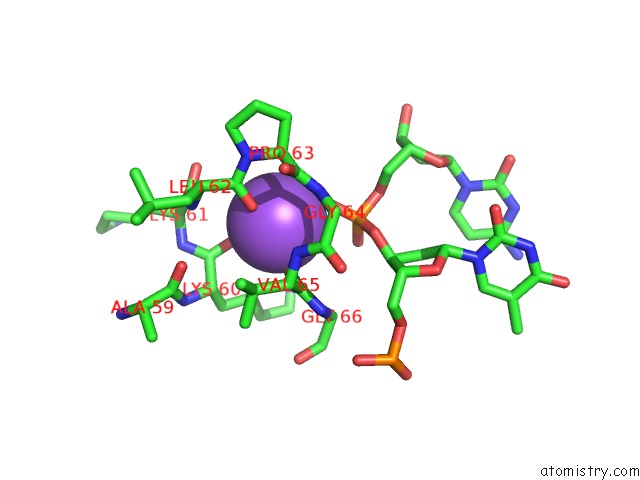
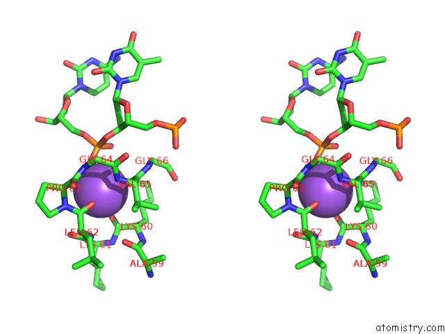
Sodium binding site 1 out of 3 in 6uol
Go back to
Sodium binding site 1 out
of 3 in the Y271G Dna Polymerase Beta Substrate Complex with A Templating Cytosine and Incoming Rgtp
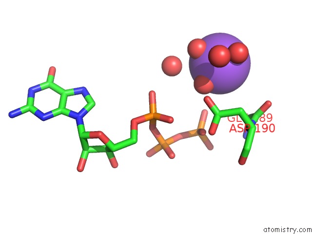
Mono view
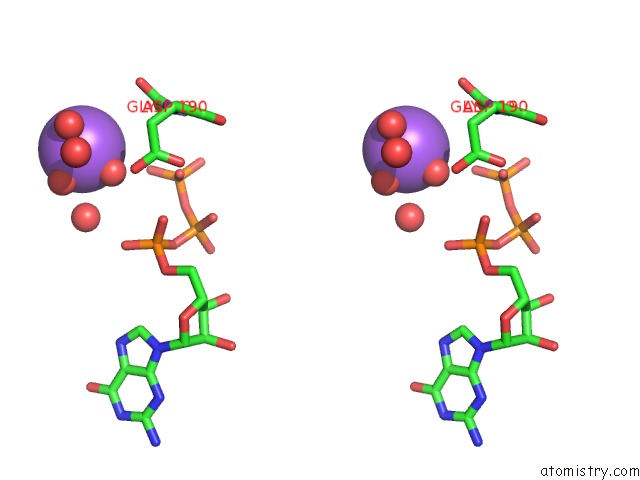
Stereo pair view
Mono view
Stereo pair view
A full contact list of Sodium with other atoms in the Na binding
site number 1 of Y271G Dna Polymerase Beta Substrate Complex with A Templating Cytosine and Incoming Rgtp within 5.0Å range:
|
Sodium binding site 2 out of 3 in 6uol
Go back to
Sodium binding site 2 out
of 3 in the Y271G Dna Polymerase Beta Substrate Complex with A Templating Cytosine and Incoming Rgtp
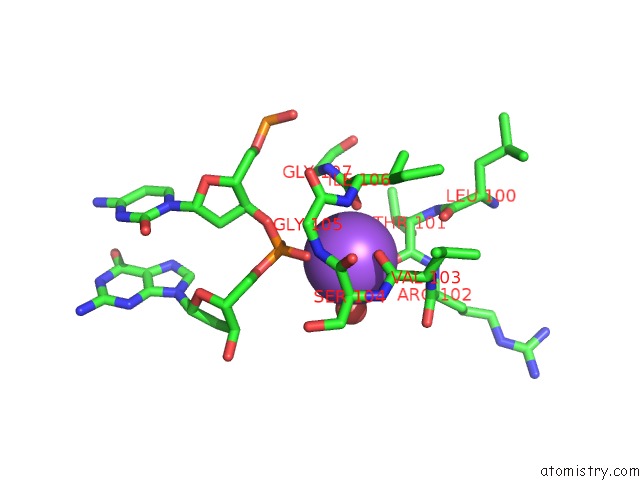
Mono view
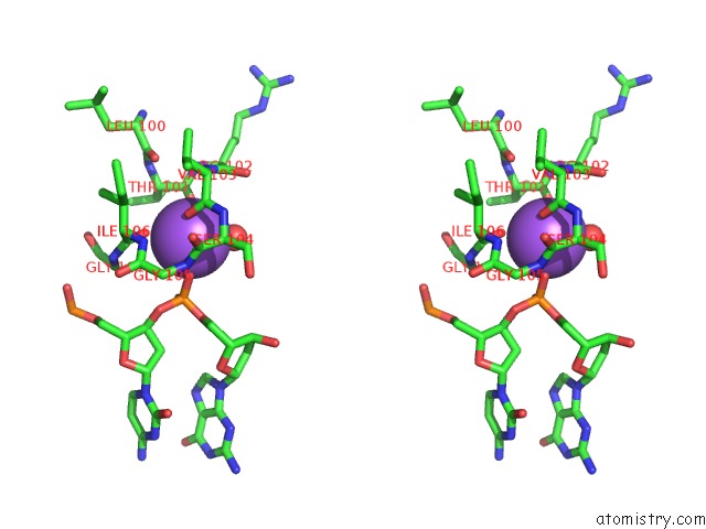
Stereo pair view
Mono view
Stereo pair view
A full contact list of Sodium with other atoms in the Na binding
site number 2 of Y271G Dna Polymerase Beta Substrate Complex with A Templating Cytosine and Incoming Rgtp within 5.0Å range:
|
Sodium binding site 3 out of 3 in 6uol
Go back to
Sodium binding site 3 out
of 3 in the Y271G Dna Polymerase Beta Substrate Complex with A Templating Cytosine and Incoming Rgtp
Mono view
Stereo pair view
Mono view
Stereo pair view
A full contact list of Sodium with other atoms in the Na binding
site number 3 of Y271G Dna Polymerase Beta Substrate Complex with A Templating Cytosine and Incoming Rgtp within 5.0Å range:
|
Reference:
M.R.Smith,
K.S.Alnajjar,
N.M.Hoitsma,
J.B.Sweasy,
B.D.Freudenthal.
Molecular Characterization of Oxidized Ribonucleotide Insertion By Human Dna Polymerase Beta J.Biol.Chem. 2020.
ISSN: ESSN 1083-351X
Page generated: Tue Oct 8 14:12:04 2024
ISSN: ESSN 1083-351X
Last articles
Zn in 9J0NZn in 9J0O
Zn in 9J0P
Zn in 9FJX
Zn in 9EKB
Zn in 9C0F
Zn in 9CAH
Zn in 9CH0
Zn in 9CH3
Zn in 9CH1