Sodium »
PDB 6u7p-6v1q »
6ucz »
Sodium in PDB 6ucz: Crystal Structure of Dihydropteroate Synthase From Anaplasma Phagocytophilum with Bound 6-Hydroxymethylpterin-Monophosphate
Enzymatic activity of Crystal Structure of Dihydropteroate Synthase From Anaplasma Phagocytophilum with Bound 6-Hydroxymethylpterin-Monophosphate
All present enzymatic activity of Crystal Structure of Dihydropteroate Synthase From Anaplasma Phagocytophilum with Bound 6-Hydroxymethylpterin-Monophosphate:
2.5.1.15;
2.5.1.15;
Protein crystallography data
The structure of Crystal Structure of Dihydropteroate Synthase From Anaplasma Phagocytophilum with Bound 6-Hydroxymethylpterin-Monophosphate, PDB code: 6ucz
was solved by
Seattle Structural Genomics Center For Infectious Disease (Ssgcid),
with X-Ray Crystallography technique. A brief refinement statistics is given in the table below:
| Resolution Low / High (Å) | 38.75 / 2.00 |
| Space group | P 1 21 1 |
| Cell size a, b, c (Å), α, β, γ (°) | 48.830, 81.480, 71.170, 90.00, 102.67, 90.00 |
| R / Rfree (%) | 16.2 / 19.8 |
Sodium Binding Sites:
The binding sites of Sodium atom in the Crystal Structure of Dihydropteroate Synthase From Anaplasma Phagocytophilum with Bound 6-Hydroxymethylpterin-Monophosphate
(pdb code 6ucz). This binding sites where shown within
5.0 Angstroms radius around Sodium atom.
In total 5 binding sites of Sodium where determined in the Crystal Structure of Dihydropteroate Synthase From Anaplasma Phagocytophilum with Bound 6-Hydroxymethylpterin-Monophosphate, PDB code: 6ucz:
Jump to Sodium binding site number: 1; 2; 3; 4; 5;
In total 5 binding sites of Sodium where determined in the Crystal Structure of Dihydropteroate Synthase From Anaplasma Phagocytophilum with Bound 6-Hydroxymethylpterin-Monophosphate, PDB code: 6ucz:
Jump to Sodium binding site number: 1; 2; 3; 4; 5;
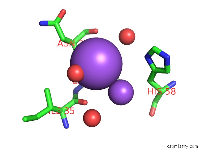
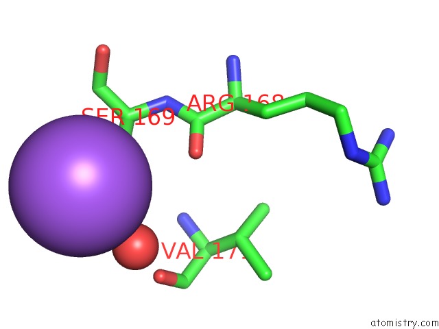
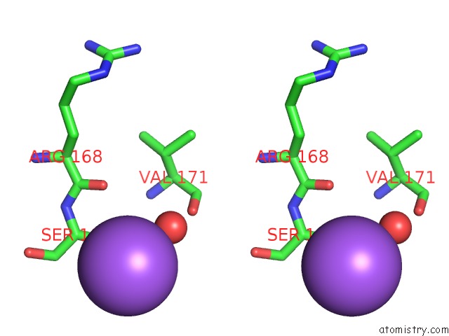
Sodium binding site 1 out of 5 in 6ucz
Go back to
Sodium binding site 1 out
of 5 in the Crystal Structure of Dihydropteroate Synthase From Anaplasma Phagocytophilum with Bound 6-Hydroxymethylpterin-Monophosphate
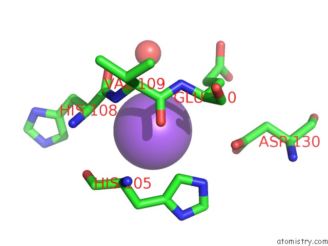
Mono view
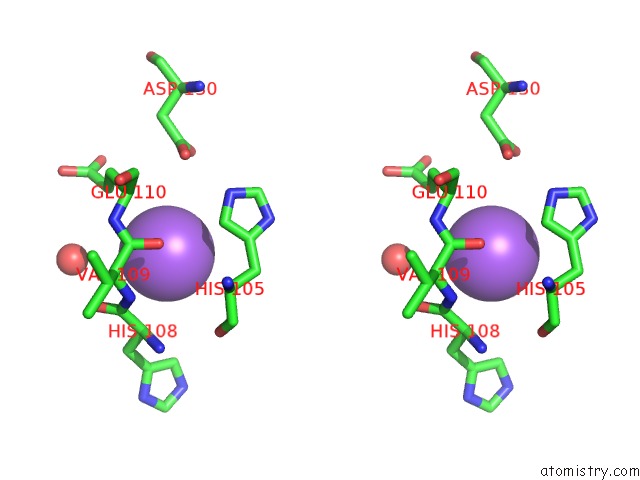
Stereo pair view
Mono view
Stereo pair view
A full contact list of Sodium with other atoms in the Na binding
site number 1 of Crystal Structure of Dihydropteroate Synthase From Anaplasma Phagocytophilum with Bound 6-Hydroxymethylpterin-Monophosphate within 5.0Å range:
|
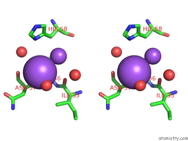
Sodium binding site 2 out of 5 in 6ucz
Go back to
Sodium binding site 2 out
of 5 in the Crystal Structure of Dihydropteroate Synthase From Anaplasma Phagocytophilum with Bound 6-Hydroxymethylpterin-Monophosphate
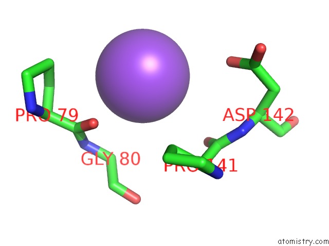
Mono view
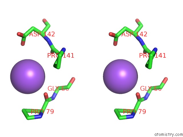
Stereo pair view
Mono view
Stereo pair view
A full contact list of Sodium with other atoms in the Na binding
site number 2 of Crystal Structure of Dihydropteroate Synthase From Anaplasma Phagocytophilum with Bound 6-Hydroxymethylpterin-Monophosphate within 5.0Å range:
|
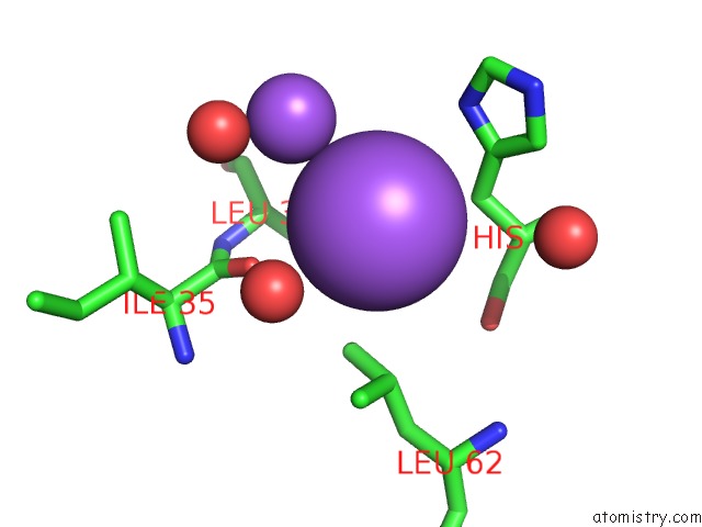
Sodium binding site 3 out of 5 in 6ucz
Go back to
Sodium binding site 3 out
of 5 in the Crystal Structure of Dihydropteroate Synthase From Anaplasma Phagocytophilum with Bound 6-Hydroxymethylpterin-Monophosphate
Mono view
Stereo pair view
Mono view
Stereo pair view
A full contact list of Sodium with other atoms in the Na binding
site number 3 of Crystal Structure of Dihydropteroate Synthase From Anaplasma Phagocytophilum with Bound 6-Hydroxymethylpterin-Monophosphate within 5.0Å range:
|
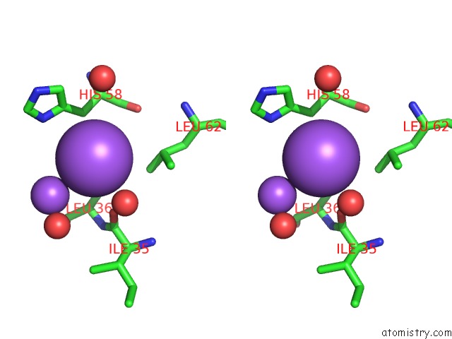
Sodium binding site 4 out of 5 in 6ucz
Go back to
Sodium binding site 4 out
of 5 in the Crystal Structure of Dihydropteroate Synthase From Anaplasma Phagocytophilum with Bound 6-Hydroxymethylpterin-Monophosphate
Mono view
Stereo pair view
Mono view
Stereo pair view
A full contact list of Sodium with other atoms in the Na binding
site number 4 of Crystal Structure of Dihydropteroate Synthase From Anaplasma Phagocytophilum with Bound 6-Hydroxymethylpterin-Monophosphate within 5.0Å range:
|
Sodium binding site 5 out of 5 in 6ucz
Go back to
Sodium binding site 5 out
of 5 in the Crystal Structure of Dihydropteroate Synthase From Anaplasma Phagocytophilum with Bound 6-Hydroxymethylpterin-Monophosphate
Mono view
Stereo pair view
Mono view
Stereo pair view
A full contact list of Sodium with other atoms in the Na binding
site number 5 of Crystal Structure of Dihydropteroate Synthase From Anaplasma Phagocytophilum with Bound 6-Hydroxymethylpterin-Monophosphate within 5.0Å range:
|
Reference:
M.J.Bolejack,
S.L.Delker,
J.Abendroth,
D.D.Lorimer,
P.S.Horanyi,
T.E.Edwards.
Crystal Structure of Dihydropteroate Synthase From Anaplasma Phagocytophilum with Bound 6-Hydroxymethylpterin-Monophosphate To Be Published.
Page generated: Mon Aug 18 07:44:30 2025
Last articles
K in 9NESK in 9PHG
K in 9NEI
K in 9NED
K in 9NEC
K in 9NEG
K in 9CWU
K in 9CVB
K in 9CVA
K in 9COM