Sodium »
PDB 6tbt-6tos »
6tod »
Sodium in PDB 6tod: Crystal Structure of the Orexin-1 Receptor in Complex with Empa
Protein crystallography data
The structure of Crystal Structure of the Orexin-1 Receptor in Complex with Empa, PDB code: 6tod
was solved by
M.Rappas,
A.Ali,
K.A.Bennett,
J.D.Brown,
S.J.Bucknell,
M.Congreve,
R.M.Cooke,
G.Cseke,
C.De Graaf,
A.S.Dore,
J.C.Errey,
A.Jazayeri,
F.H.Marshall,
J.S.Mason,
R.Mould,
J.C.Patel,
B.G.Tehan,
M.Weir,
J.A.Christopher,
with X-Ray Crystallography technique. A brief refinement statistics is given in the table below:
| Resolution Low / High (Å) | 30.62 / 2.11 |
| Space group | I 1 2 1 |
| Cell size a, b, c (Å), α, β, γ (°) | 57.907, 158.891, 182.354, 90.00, 95.77, 90.00 |
| R / Rfree (%) | 18.8 / 20.8 |
Sodium Binding Sites:
The binding sites of Sodium atom in the Crystal Structure of the Orexin-1 Receptor in Complex with Empa
(pdb code 6tod). This binding sites where shown within
5.0 Angstroms radius around Sodium atom.
In total 2 binding sites of Sodium where determined in the Crystal Structure of the Orexin-1 Receptor in Complex with Empa, PDB code: 6tod:
Jump to Sodium binding site number: 1; 2;
In total 2 binding sites of Sodium where determined in the Crystal Structure of the Orexin-1 Receptor in Complex with Empa, PDB code: 6tod:
Jump to Sodium binding site number: 1; 2;
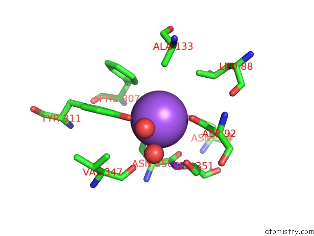
Sodium binding site 1 out of 2 in 6tod
Go back to
Sodium binding site 1 out
of 2 in the Crystal Structure of the Orexin-1 Receptor in Complex with Empa
Mono view
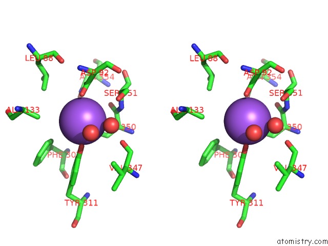
Stereo pair view
Mono view
Stereo pair view
A full contact list of Sodium with other atoms in the Na binding
site number 1 of Crystal Structure of the Orexin-1 Receptor in Complex with Empa within 5.0Å range:
|
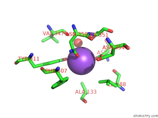
Sodium binding site 2 out of 2 in 6tod
Go back to
Sodium binding site 2 out
of 2 in the Crystal Structure of the Orexin-1 Receptor in Complex with Empa
Mono view
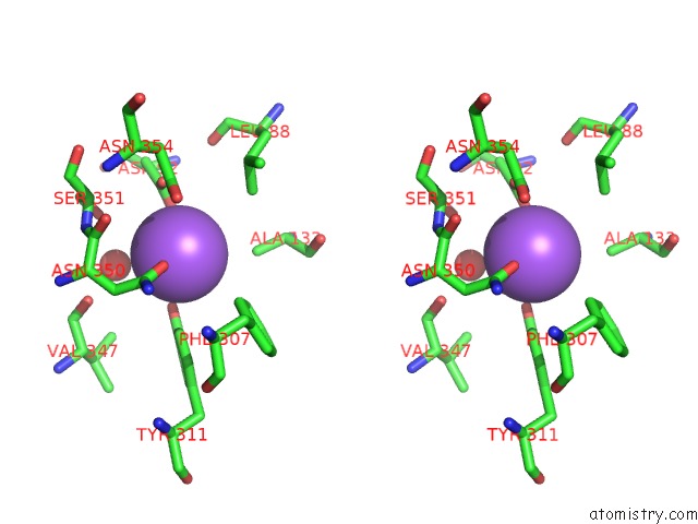
Stereo pair view
Mono view
Stereo pair view
A full contact list of Sodium with other atoms in the Na binding
site number 2 of Crystal Structure of the Orexin-1 Receptor in Complex with Empa within 5.0Å range:
|
Reference:
M.Rappas,
A.Ali,
K.A.Bennett,
J.D.Brown,
S.J.Bucknell,
M.Congreve,
R.M.Cooke,
G.Cseke,
C.De Graaf,
A.S.Dore,
J.C.Errey,
A.Jazayeri,
F.H.Marshall,
J.S.Mason,
R.Mould,
J.C.Patel,
B.Tehan,
M.Weir,
J.A.Christopher.
Comparison of Orexin 1 and Orexin 2 Ligand Binding Modes Using X-Ray Crystallography and Computational Analysis. J.Med.Chem. 2019.
ISSN: ISSN 0022-2623
PubMed: 31860301
DOI: 10.1021/ACS.JMEDCHEM.9B01787
Page generated: Tue Oct 8 14:00:09 2024
ISSN: ISSN 0022-2623
PubMed: 31860301
DOI: 10.1021/ACS.JMEDCHEM.9B01787
Last articles
Zn in 9J0NZn in 9J0O
Zn in 9J0P
Zn in 9FJX
Zn in 9EKB
Zn in 9C0F
Zn in 9CAH
Zn in 9CH0
Zn in 9CH3
Zn in 9CH1