Sodium »
PDB 6qtp-6r7k »
6r6k »
Sodium in PDB 6r6k: Structure of A Fpvc Mutant From Pseudomonas Aeruginosa
Protein crystallography data
The structure of Structure of A Fpvc Mutant From Pseudomonas Aeruginosa, PDB code: 6r6k
was solved by
S.Morera,
A.Vigouroux,
with X-Ray Crystallography technique. A brief refinement statistics is given in the table below:
| Resolution Low / High (Å) | 43.74 / 2.10 |
| Space group | C 1 2 1 |
| Cell size a, b, c (Å), α, β, γ (°) | 155.270, 53.560, 84.390, 90.00, 93.16, 90.00 |
| R / Rfree (%) | 19.4 / 23.4 |
Sodium Binding Sites:
The binding sites of Sodium atom in the Structure of A Fpvc Mutant From Pseudomonas Aeruginosa
(pdb code 6r6k). This binding sites where shown within
5.0 Angstroms radius around Sodium atom.
In total 6 binding sites of Sodium where determined in the Structure of A Fpvc Mutant From Pseudomonas Aeruginosa, PDB code: 6r6k:
Jump to Sodium binding site number: 1; 2; 3; 4; 5; 6;
In total 6 binding sites of Sodium where determined in the Structure of A Fpvc Mutant From Pseudomonas Aeruginosa, PDB code: 6r6k:
Jump to Sodium binding site number: 1; 2; 3; 4; 5; 6;
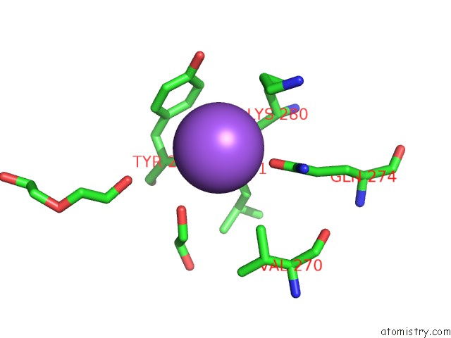
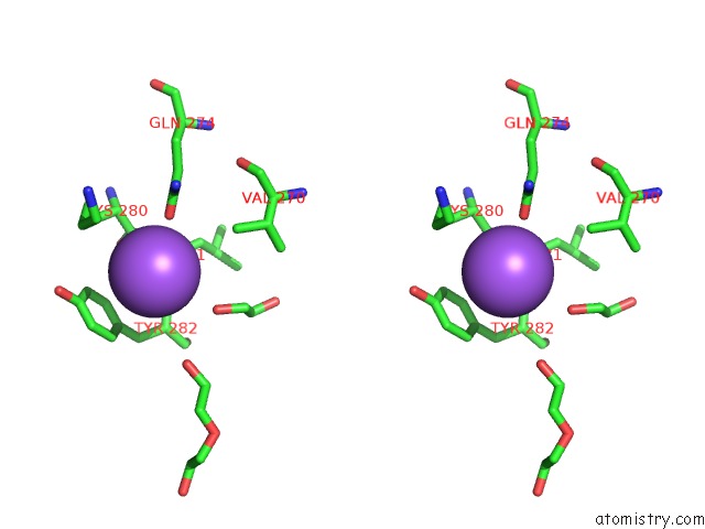
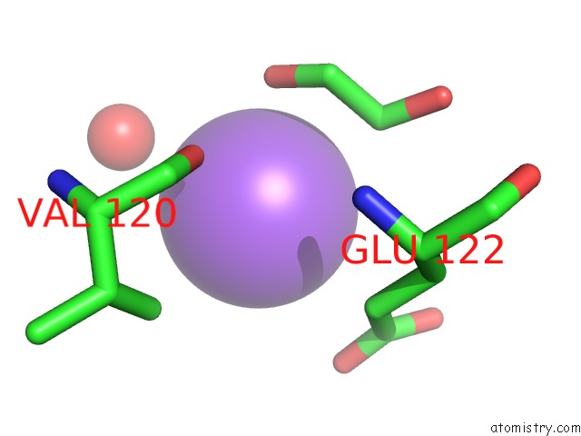
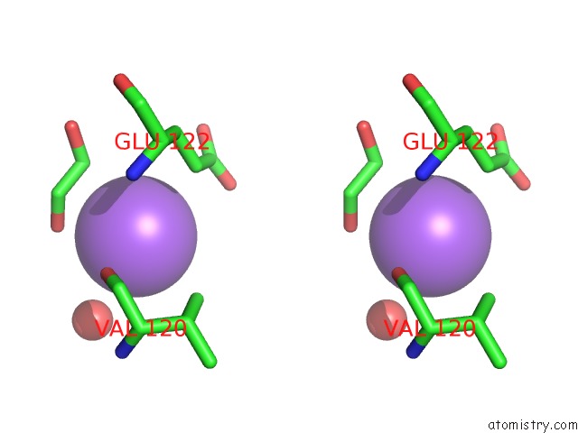
Sodium binding site 1 out of 6 in 6r6k
Go back to
Sodium binding site 1 out
of 6 in the Structure of A Fpvc Mutant From Pseudomonas Aeruginosa
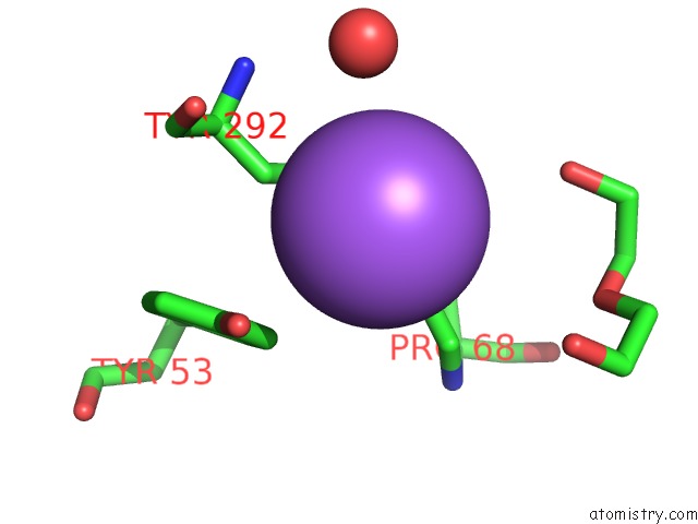
Mono view
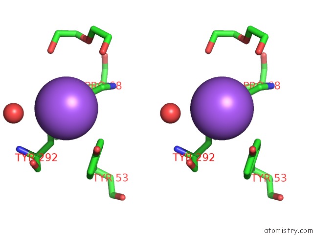
Stereo pair view
Mono view
Stereo pair view
A full contact list of Sodium with other atoms in the Na binding
site number 1 of Structure of A Fpvc Mutant From Pseudomonas Aeruginosa within 5.0Å range:
|
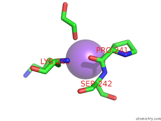
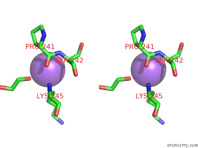
Sodium binding site 2 out of 6 in 6r6k
Go back to
Sodium binding site 2 out
of 6 in the Structure of A Fpvc Mutant From Pseudomonas Aeruginosa
Mono view
Stereo pair view
Mono view
Stereo pair view
A full contact list of Sodium with other atoms in the Na binding
site number 2 of Structure of A Fpvc Mutant From Pseudomonas Aeruginosa within 5.0Å range:
|
Sodium binding site 3 out of 6 in 6r6k
Go back to
Sodium binding site 3 out
of 6 in the Structure of A Fpvc Mutant From Pseudomonas Aeruginosa
Mono view
Stereo pair view
Mono view
Stereo pair view
A full contact list of Sodium with other atoms in the Na binding
site number 3 of Structure of A Fpvc Mutant From Pseudomonas Aeruginosa within 5.0Å range:
|
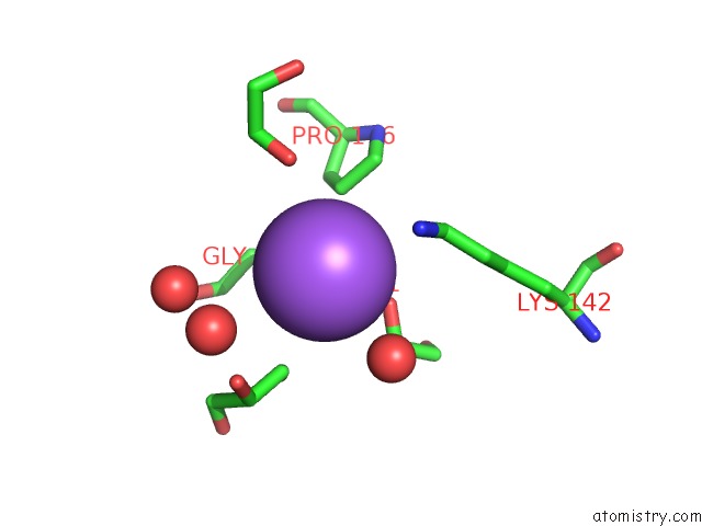
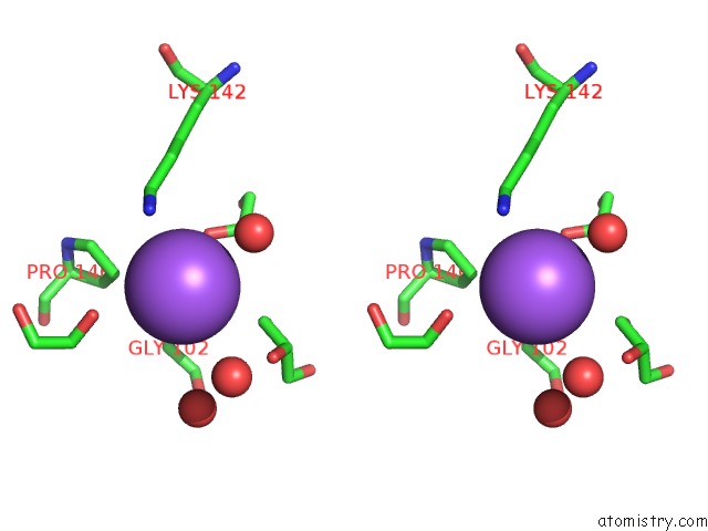
Sodium binding site 4 out of 6 in 6r6k
Go back to
Sodium binding site 4 out
of 6 in the Structure of A Fpvc Mutant From Pseudomonas Aeruginosa
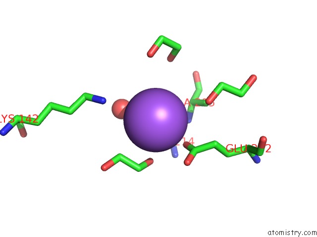
Mono view
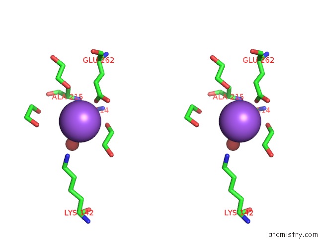
Stereo pair view
Mono view
Stereo pair view
A full contact list of Sodium with other atoms in the Na binding
site number 4 of Structure of A Fpvc Mutant From Pseudomonas Aeruginosa within 5.0Å range:
|
Sodium binding site 5 out of 6 in 6r6k
Go back to
Sodium binding site 5 out
of 6 in the Structure of A Fpvc Mutant From Pseudomonas Aeruginosa
Mono view
Stereo pair view
Mono view
Stereo pair view
A full contact list of Sodium with other atoms in the Na binding
site number 5 of Structure of A Fpvc Mutant From Pseudomonas Aeruginosa within 5.0Å range:
|
Sodium binding site 6 out of 6 in 6r6k
Go back to
Sodium binding site 6 out
of 6 in the Structure of A Fpvc Mutant From Pseudomonas Aeruginosa
Mono view
Stereo pair view
Mono view
Stereo pair view
A full contact list of Sodium with other atoms in the Na binding
site number 6 of Structure of A Fpvc Mutant From Pseudomonas Aeruginosa within 5.0Å range:
|
Reference:
A.Vigouroux,
M.Aumont-Nicaise,
A.Boussac,
L.Marty,
L.Lo Bello,
P.Legrand,
K.Brillet,
I.J.Schalk,
S.Morera.
A Unique Ferrous Iron Binding Mode Is Associated with Large Conformational Changes For the Transport Protein Fpvc of Pseudomonas Aeruginosa. Febs J. 2019.
ISSN: ISSN 1742-464X
PubMed: 31318478
DOI: 10.1111/FEBS.15004
Page generated: Tue Oct 8 13:04:15 2024
ISSN: ISSN 1742-464X
PubMed: 31318478
DOI: 10.1111/FEBS.15004
Last articles
Ca in 5O25Ca in 5O1U
Ca in 5O0S
Ca in 5NZE
Ca in 5NZ4
Ca in 5NWE
Ca in 5NYY
Ca in 5NXL
Ca in 5NXU
Ca in 5NXR