Sodium »
PDB 6oor-6p8m »
6ozl »
Sodium in PDB 6ozl: Crystal Structure of Mus Musculus (Mm) Endonuclease V in Complex with A 23MER Rna Oligo Containing An Inosine After A 2 Min Soak in MN2+
Protein crystallography data
The structure of Crystal Structure of Mus Musculus (Mm) Endonuclease V in Complex with A 23MER Rna Oligo Containing An Inosine After A 2 Min Soak in MN2+, PDB code: 6ozl
was solved by
N.L.Samara,
W.Yang,
with X-Ray Crystallography technique. A brief refinement statistics is given in the table below:
| Resolution Low / High (Å) | 35.82 / 2.10 |
| Space group | P 21 21 21 |
| Cell size a, b, c (Å), α, β, γ (°) | 73.621, 72.519, 155.764, 90.00, 90.00, 90.00 |
| R / Rfree (%) | 17.6 / 21.7 |
Other elements in 6ozl:
The structure of Crystal Structure of Mus Musculus (Mm) Endonuclease V in Complex with A 23MER Rna Oligo Containing An Inosine After A 2 Min Soak in MN2+ also contains other interesting chemical elements:
| Manganese | (Mn) | 4 atoms |
Sodium Binding Sites:
The binding sites of Sodium atom in the Crystal Structure of Mus Musculus (Mm) Endonuclease V in Complex with A 23MER Rna Oligo Containing An Inosine After A 2 Min Soak in MN2+
(pdb code 6ozl). This binding sites where shown within
5.0 Angstroms radius around Sodium atom.
In total only one binding site of Sodium was determined in the Crystal Structure of Mus Musculus (Mm) Endonuclease V in Complex with A 23MER Rna Oligo Containing An Inosine After A 2 Min Soak in MN2+, PDB code: 6ozl:
In total only one binding site of Sodium was determined in the Crystal Structure of Mus Musculus (Mm) Endonuclease V in Complex with A 23MER Rna Oligo Containing An Inosine After A 2 Min Soak in MN2+, PDB code: 6ozl:
Sodium binding site 1 out of 1 in 6ozl
Go back to
Sodium binding site 1 out
of 1 in the Crystal Structure of Mus Musculus (Mm) Endonuclease V in Complex with A 23MER Rna Oligo Containing An Inosine After A 2 Min Soak in MN2+
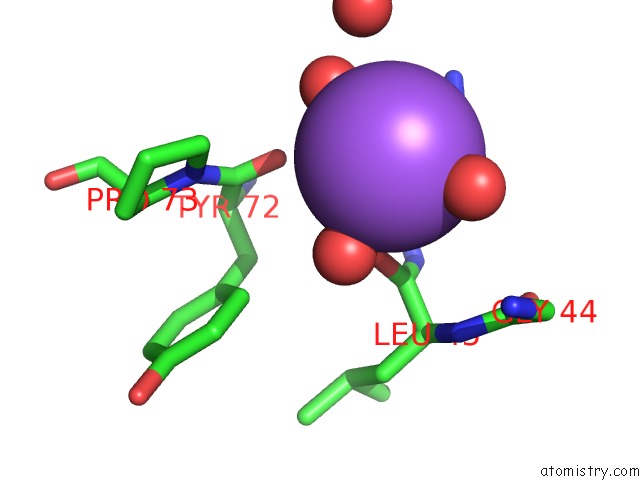
Mono view
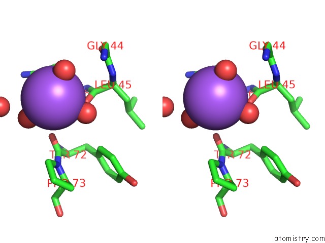
Stereo pair view
Mono view
Stereo pair view
A full contact list of Sodium with other atoms in the Na binding
site number 1 of Crystal Structure of Mus Musculus (Mm) Endonuclease V in Complex with A 23MER Rna Oligo Containing An Inosine After A 2 Min Soak in MN2+ within 5.0Å range:
|
Reference:
J.Wu,
N.L.Samara,
I.Kuraoka,
W.Yang.
Evolution of Inosine-Specific Endonuclease V From Bacterial Dnase to Eukaryotic Rnase. Mol.Cell V. 76 44 2019.
ISSN: ISSN 1097-2765
PubMed: 31444105
DOI: 10.1016/J.MOLCEL.2019.06.046
Page generated: Tue Oct 8 12:38:47 2024
ISSN: ISSN 1097-2765
PubMed: 31444105
DOI: 10.1016/J.MOLCEL.2019.06.046
Last articles
Zn in 9J0NZn in 9J0O
Zn in 9J0P
Zn in 9FJX
Zn in 9EKB
Zn in 9C0F
Zn in 9CAH
Zn in 9CH0
Zn in 9CH3
Zn in 9CH1