Sodium »
PDB 6nib-6nxy »
6nuf »
Sodium in PDB 6nuf: Structure of Calcineurin in Complex with NHE1 Peptide
Enzymatic activity of Structure of Calcineurin in Complex with NHE1 Peptide
All present enzymatic activity of Structure of Calcineurin in Complex with NHE1 Peptide:
3.1.3.16;
3.1.3.16;
Protein crystallography data
The structure of Structure of Calcineurin in Complex with NHE1 Peptide, PDB code: 6nuf
was solved by
X.Wang,
R.Page,
W.Peti,
with X-Ray Crystallography technique. A brief refinement statistics is given in the table below:
| Resolution Low / High (Å) | 16.13 / 1.90 |
| Space group | C 2 2 21 |
| Cell size a, b, c (Å), α, β, γ (°) | 79.477, 126.808, 127.431, 90.00, 90.00, 90.00 |
| R / Rfree (%) | 16.9 / 20.9 |
Other elements in 6nuf:
The structure of Structure of Calcineurin in Complex with NHE1 Peptide also contains other interesting chemical elements:
| Zinc | (Zn) | 1 atom |
| Iron | (Fe) | 1 atom |
| Calcium | (Ca) | 4 atoms |
Sodium Binding Sites:
The binding sites of Sodium atom in the Structure of Calcineurin in Complex with NHE1 Peptide
(pdb code 6nuf). This binding sites where shown within
5.0 Angstroms radius around Sodium atom.
In total only one binding site of Sodium was determined in the Structure of Calcineurin in Complex with NHE1 Peptide, PDB code: 6nuf:
In total only one binding site of Sodium was determined in the Structure of Calcineurin in Complex with NHE1 Peptide, PDB code: 6nuf:
Sodium binding site 1 out of 1 in 6nuf
Go back to
Sodium binding site 1 out
of 1 in the Structure of Calcineurin in Complex with NHE1 Peptide
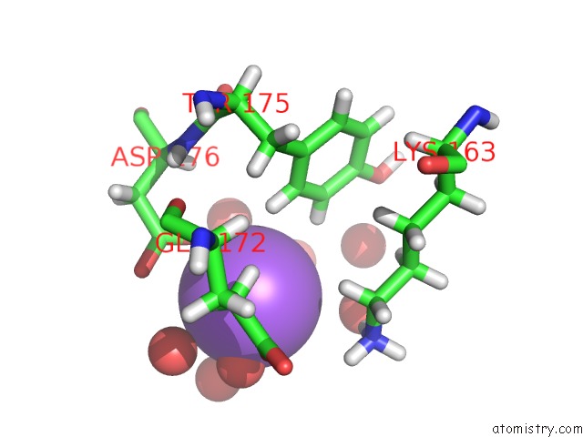
Mono view
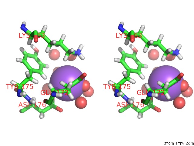
Stereo pair view
Mono view
Stereo pair view
A full contact list of Sodium with other atoms in the Na binding
site number 1 of Structure of Calcineurin in Complex with NHE1 Peptide within 5.0Å range:
|
Reference:
R.Hendus-Altenburger,
X.Wang,
L.M.Sjogaard-Frich,
E.Pedraz-Cuesta,
S.R.Sheftic,
A.H.Bendsoe,
R.Page,
B.B.Kragelund,
S.F.Pedersen,
W.Peti.
Molecular Basis For the Binding and Selective Dephosphorylation of Na+/H+Exchanger 1 By Calcineurin. Nat Commun V. 10 3489 2019.
ISSN: ESSN 2041-1723
PubMed: 31375679
DOI: 10.1038/S41467-019-11391-7
Page generated: Tue Oct 8 12:22:05 2024
ISSN: ESSN 2041-1723
PubMed: 31375679
DOI: 10.1038/S41467-019-11391-7
Last articles
Cl in 7ZWXCl in 7ZWT
Cl in 7ZWU
Cl in 7ZW2
Cl in 7ZWS
Cl in 7ZWO
Cl in 7ZW7
Cl in 7ZVO
Cl in 7ZV3
Cl in 7ZVH