Sodium »
PDB 6lh7-6mec »
6m38 »
Sodium in PDB 6m38: X-Ray Structure of A Drosophila Dopamine Transporter with Subsiteb Mutations (D121G/S426M) in S-Duloxetine Bound Form
Protein crystallography data
The structure of X-Ray Structure of A Drosophila Dopamine Transporter with Subsiteb Mutations (D121G/S426M) in S-Duloxetine Bound Form, PDB code: 6m38
was solved by
P.Shabareesh,
A.K.Mallela,
D.Joseph,
A.Penmatsa,
with X-Ray Crystallography technique. A brief refinement statistics is given in the table below:
| Resolution Low / High (Å) | 48.64 / 3.00 |
| Space group | P 21 21 21 |
| Cell size a, b, c (Å), α, β, γ (°) | 97.288, 140.641, 167.818, 90, 90, 90 |
| R / Rfree (%) | 21.5 / 24.4 |
Other elements in 6m38:
The structure of X-Ray Structure of A Drosophila Dopamine Transporter with Subsiteb Mutations (D121G/S426M) in S-Duloxetine Bound Form also contains other interesting chemical elements:
| Chlorine | (Cl) | 1 atom |
Sodium Binding Sites:
The binding sites of Sodium atom in the X-Ray Structure of A Drosophila Dopamine Transporter with Subsiteb Mutations (D121G/S426M) in S-Duloxetine Bound Form
(pdb code 6m38). This binding sites where shown within
5.0 Angstroms radius around Sodium atom.
In total 2 binding sites of Sodium where determined in the X-Ray Structure of A Drosophila Dopamine Transporter with Subsiteb Mutations (D121G/S426M) in S-Duloxetine Bound Form, PDB code: 6m38:
Jump to Sodium binding site number: 1; 2;
In total 2 binding sites of Sodium where determined in the X-Ray Structure of A Drosophila Dopamine Transporter with Subsiteb Mutations (D121G/S426M) in S-Duloxetine Bound Form, PDB code: 6m38:
Jump to Sodium binding site number: 1; 2;
Sodium binding site 1 out of 2 in 6m38
Go back to
Sodium binding site 1 out
of 2 in the X-Ray Structure of A Drosophila Dopamine Transporter with Subsiteb Mutations (D121G/S426M) in S-Duloxetine Bound Form
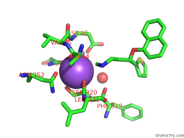
Mono view
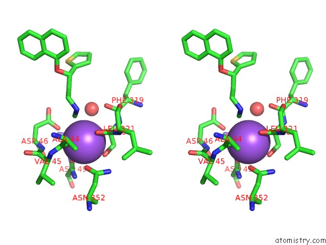
Stereo pair view
Mono view
Stereo pair view
A full contact list of Sodium with other atoms in the Na binding
site number 1 of X-Ray Structure of A Drosophila Dopamine Transporter with Subsiteb Mutations (D121G/S426M) in S-Duloxetine Bound Form within 5.0Å range:
|
Sodium binding site 2 out of 2 in 6m38
Go back to
Sodium binding site 2 out
of 2 in the X-Ray Structure of A Drosophila Dopamine Transporter with Subsiteb Mutations (D121G/S426M) in S-Duloxetine Bound Form
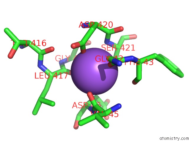
Mono view
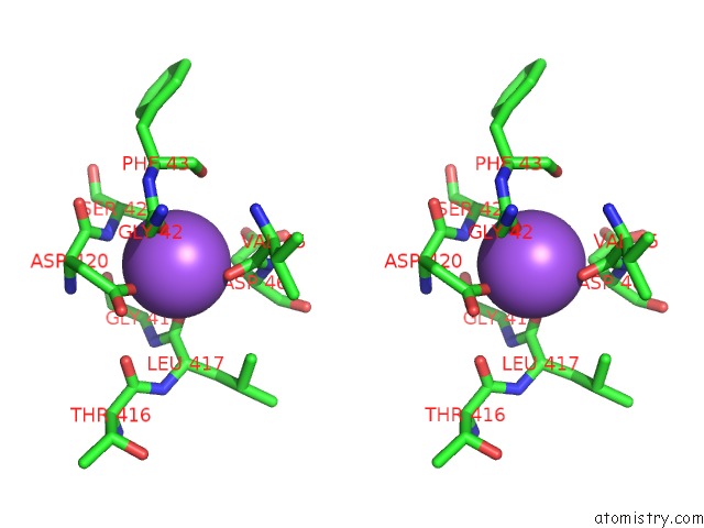
Stereo pair view
Mono view
Stereo pair view
A full contact list of Sodium with other atoms in the Na binding
site number 2 of X-Ray Structure of A Drosophila Dopamine Transporter with Subsiteb Mutations (D121G/S426M) in S-Duloxetine Bound Form within 5.0Å range:
|
Reference:
P.Shabareesh,
A.K.Mallela,
D.Joseph.
Structural Basis of Norepinephrine Recognition and Transport Inhibition in Neurotransmitter Transporters To Be Published.
Page generated: Mon Aug 18 05:54:43 2025
Last articles
Na in 6Z50Na in 6YU8
Na in 6Z31
Na in 6Z3X
Na in 6Z19
Na in 6Z0L
Na in 6YZS
Na in 6Z02
Na in 6YZ2
Na in 6YZH