Sodium »
PDB 6g3r-6gn7 »
6gn7 »
Sodium in PDB 6gn7: X-Ray Structure of the Complex Between Human Alpha Thrombin and NU172, A Duplex/Quadruplex 26-Mer Dna Aptamer, in the Presence of Sodium Ions.
Enzymatic activity of X-Ray Structure of the Complex Between Human Alpha Thrombin and NU172, A Duplex/Quadruplex 26-Mer Dna Aptamer, in the Presence of Sodium Ions.
All present enzymatic activity of X-Ray Structure of the Complex Between Human Alpha Thrombin and NU172, A Duplex/Quadruplex 26-Mer Dna Aptamer, in the Presence of Sodium Ions.:
3.4.21.5;
3.4.21.5;
Protein crystallography data
The structure of X-Ray Structure of the Complex Between Human Alpha Thrombin and NU172, A Duplex/Quadruplex 26-Mer Dna Aptamer, in the Presence of Sodium Ions., PDB code: 6gn7
was solved by
R.Troisi,
I.Russo Krauss,
F.Sica,
with X-Ray Crystallography technique. A brief refinement statistics is given in the table below:
| Resolution Low / High (Å) | 104.51 / 2.80 |
| Space group | I 2 2 2 |
| Cell size a, b, c (Å), α, β, γ (°) | 67.330, 120.690, 208.940, 90.00, 90.00, 90.00 |
| R / Rfree (%) | 15.9 / 20.3 |
Sodium Binding Sites:
The binding sites of Sodium atom in the X-Ray Structure of the Complex Between Human Alpha Thrombin and NU172, A Duplex/Quadruplex 26-Mer Dna Aptamer, in the Presence of Sodium Ions.
(pdb code 6gn7). This binding sites where shown within
5.0 Angstroms radius around Sodium atom.
In total 2 binding sites of Sodium where determined in the X-Ray Structure of the Complex Between Human Alpha Thrombin and NU172, A Duplex/Quadruplex 26-Mer Dna Aptamer, in the Presence of Sodium Ions., PDB code: 6gn7:
Jump to Sodium binding site number: 1; 2;
In total 2 binding sites of Sodium where determined in the X-Ray Structure of the Complex Between Human Alpha Thrombin and NU172, A Duplex/Quadruplex 26-Mer Dna Aptamer, in the Presence of Sodium Ions., PDB code: 6gn7:
Jump to Sodium binding site number: 1; 2;
Sodium binding site 1 out of 2 in 6gn7
Go back to
Sodium binding site 1 out
of 2 in the X-Ray Structure of the Complex Between Human Alpha Thrombin and NU172, A Duplex/Quadruplex 26-Mer Dna Aptamer, in the Presence of Sodium Ions.
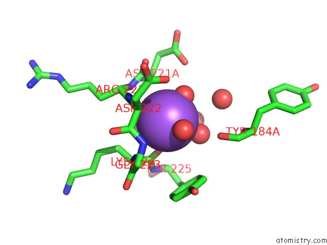
Mono view
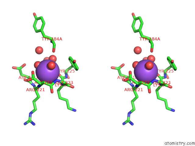
Stereo pair view
Mono view
Stereo pair view
A full contact list of Sodium with other atoms in the Na binding
site number 1 of X-Ray Structure of the Complex Between Human Alpha Thrombin and NU172, A Duplex/Quadruplex 26-Mer Dna Aptamer, in the Presence of Sodium Ions. within 5.0Å range:
|
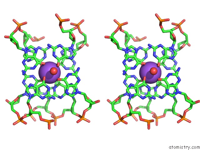
Sodium binding site 2 out of 2 in 6gn7
Go back to
Sodium binding site 2 out
of 2 in the X-Ray Structure of the Complex Between Human Alpha Thrombin and NU172, A Duplex/Quadruplex 26-Mer Dna Aptamer, in the Presence of Sodium Ions.
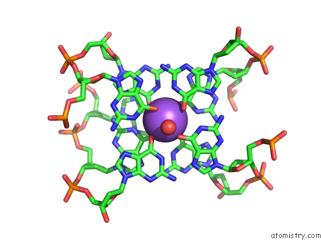
Mono view
Stereo pair view
Mono view
Stereo pair view
A full contact list of Sodium with other atoms in the Na binding
site number 2 of X-Ray Structure of the Complex Between Human Alpha Thrombin and NU172, A Duplex/Quadruplex 26-Mer Dna Aptamer, in the Presence of Sodium Ions. within 5.0Å range:
|
Reference:
R.Troisi,
V.Napolitano,
V.Spiridonova,
I.Russo Krauss,
F.Sica.
Several Structural Motifs Cooperate in Determining the Highly Effective Anti-Thrombin Activity of NU172 Aptamer. Nucleic Acids Res. V. 46 12177 2018.
ISSN: ESSN 1362-4962
PubMed: 30357392
DOI: 10.1093/NAR/GKY990
Page generated: Tue Oct 8 09:02:44 2024
ISSN: ESSN 1362-4962
PubMed: 30357392
DOI: 10.1093/NAR/GKY990
Last articles
Zn in 9J0NZn in 9J0O
Zn in 9J0P
Zn in 9FJX
Zn in 9EKB
Zn in 9C0F
Zn in 9CAH
Zn in 9CH0
Zn in 9CH3
Zn in 9CH1