Sodium »
PDB 6g3r-6gn7 »
6gbn »
Sodium in PDB 6gbn: Crystal Structure of S-Adenosyl-L-Homocysteine Hydrolase From Cytophaga Hutchinsonii in Complex with Adenosine
Enzymatic activity of Crystal Structure of S-Adenosyl-L-Homocysteine Hydrolase From Cytophaga Hutchinsonii in Complex with Adenosine
All present enzymatic activity of Crystal Structure of S-Adenosyl-L-Homocysteine Hydrolase From Cytophaga Hutchinsonii in Complex with Adenosine:
3.3.1.1;
3.3.1.1;
Protein crystallography data
The structure of Crystal Structure of S-Adenosyl-L-Homocysteine Hydrolase From Cytophaga Hutchinsonii in Complex with Adenosine, PDB code: 6gbn
was solved by
J.Czyrko,
M.Jaskolski,
K.Brzezinski,
with X-Ray Crystallography technique. A brief refinement statistics is given in the table below:
| Resolution Low / High (Å) | 48.14 / 2.09 |
| Space group | P 21 21 2 |
| Cell size a, b, c (Å), α, β, γ (°) | 96.275, 102.445, 188.919, 90.00, 90.00, 90.00 |
| R / Rfree (%) | 21 / 23.1 |
Sodium Binding Sites:
The binding sites of Sodium atom in the Crystal Structure of S-Adenosyl-L-Homocysteine Hydrolase From Cytophaga Hutchinsonii in Complex with Adenosine
(pdb code 6gbn). This binding sites where shown within
5.0 Angstroms radius around Sodium atom.
In total 4 binding sites of Sodium where determined in the Crystal Structure of S-Adenosyl-L-Homocysteine Hydrolase From Cytophaga Hutchinsonii in Complex with Adenosine, PDB code: 6gbn:
Jump to Sodium binding site number: 1; 2; 3; 4;
In total 4 binding sites of Sodium where determined in the Crystal Structure of S-Adenosyl-L-Homocysteine Hydrolase From Cytophaga Hutchinsonii in Complex with Adenosine, PDB code: 6gbn:
Jump to Sodium binding site number: 1; 2; 3; 4;
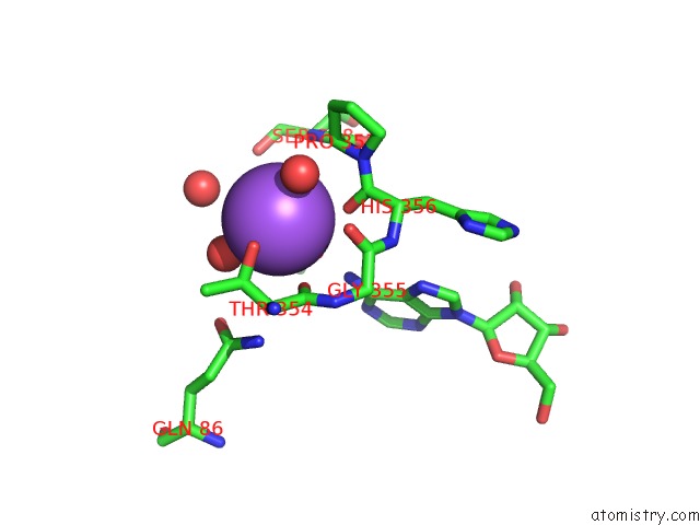
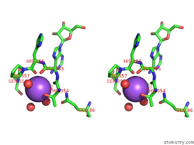
Sodium binding site 1 out of 4 in 6gbn
Go back to
Sodium binding site 1 out
of 4 in the Crystal Structure of S-Adenosyl-L-Homocysteine Hydrolase From Cytophaga Hutchinsonii in Complex with Adenosine
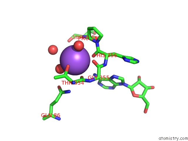
Mono view
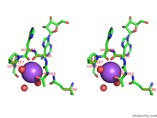
Stereo pair view
Mono view
Stereo pair view
A full contact list of Sodium with other atoms in the Na binding
site number 1 of Crystal Structure of S-Adenosyl-L-Homocysteine Hydrolase From Cytophaga Hutchinsonii in Complex with Adenosine within 5.0Å range:
|
Sodium binding site 2 out of 4 in 6gbn
Go back to
Sodium binding site 2 out
of 4 in the Crystal Structure of S-Adenosyl-L-Homocysteine Hydrolase From Cytophaga Hutchinsonii in Complex with Adenosine
Mono view
Stereo pair view
Mono view
Stereo pair view
A full contact list of Sodium with other atoms in the Na binding
site number 2 of Crystal Structure of S-Adenosyl-L-Homocysteine Hydrolase From Cytophaga Hutchinsonii in Complex with Adenosine within 5.0Å range:
|
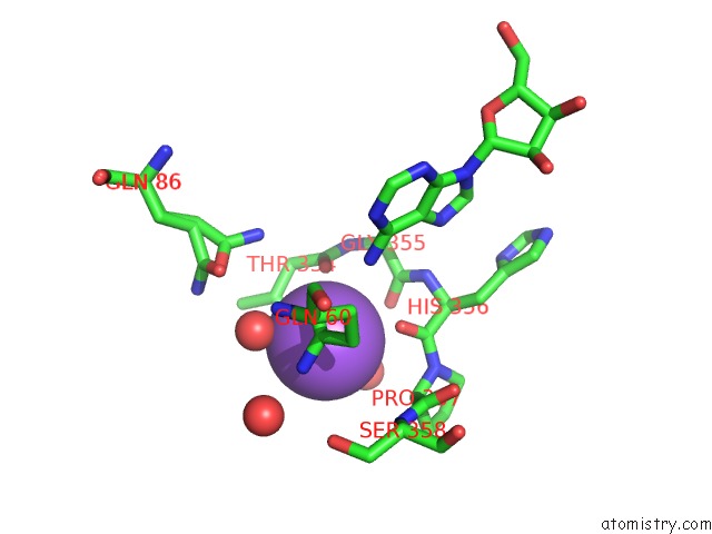
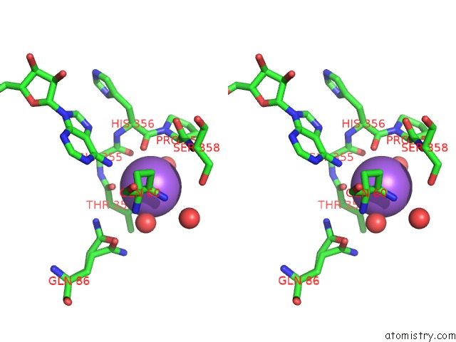
Sodium binding site 3 out of 4 in 6gbn
Go back to
Sodium binding site 3 out
of 4 in the Crystal Structure of S-Adenosyl-L-Homocysteine Hydrolase From Cytophaga Hutchinsonii in Complex with Adenosine
Mono view
Stereo pair view
Mono view
Stereo pair view
A full contact list of Sodium with other atoms in the Na binding
site number 3 of Crystal Structure of S-Adenosyl-L-Homocysteine Hydrolase From Cytophaga Hutchinsonii in Complex with Adenosine within 5.0Å range:
|
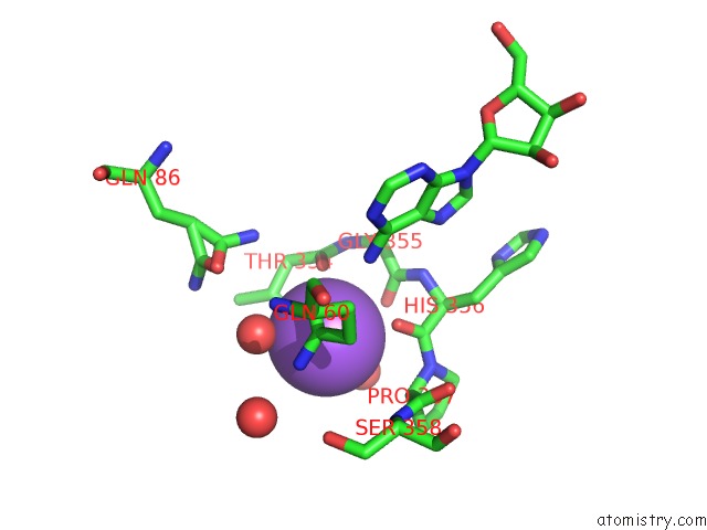
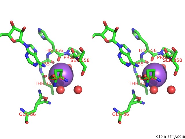
Sodium binding site 4 out of 4 in 6gbn
Go back to
Sodium binding site 4 out
of 4 in the Crystal Structure of S-Adenosyl-L-Homocysteine Hydrolase From Cytophaga Hutchinsonii in Complex with Adenosine
Mono view
Stereo pair view
Mono view
Stereo pair view
A full contact list of Sodium with other atoms in the Na binding
site number 4 of Crystal Structure of S-Adenosyl-L-Homocysteine Hydrolase From Cytophaga Hutchinsonii in Complex with Adenosine within 5.0Å range:
|
Reference:
J.Czyrko,
M.Jaskolski,
K.Brzezinski.
Crystal Structure of S-Adenosyl-L-Homocysteine Hydrolase From Cytophaga Hutchinsonii, A Case of Combination of Crystallographic and Non-Crystallographic Symmetry. Croatica Chemica Acta V. 91 2018.
ISSN: ISSN 0011-1643
DOI: 10.5562/CCA3345
Page generated: Tue Oct 8 08:59:31 2024
ISSN: ISSN 0011-1643
DOI: 10.5562/CCA3345
Last articles
Zn in 9J0NZn in 9J0O
Zn in 9J0P
Zn in 9FJX
Zn in 9EKB
Zn in 9C0F
Zn in 9CAH
Zn in 9CH0
Zn in 9CH3
Zn in 9CH1