Sodium »
PDB 6g3r-6gn7 »
6g7o »
Sodium in PDB 6g7o: Crystal Structure of Human Alkaline Ceramidase 3 (ACER3) at 2.7 Angstrom Resolution
Protein crystallography data
The structure of Crystal Structure of Human Alkaline Ceramidase 3 (ACER3) at 2.7 Angstrom Resolution, PDB code: 6g7o
was solved by
C.Leyrat,
I.Vasiliauskaite-Brooks,
R.D.Healey,
R.Sounier,
C.Grison,
F.Hoh,
S.Basu,
S.Granier,
with X-Ray Crystallography technique. A brief refinement statistics is given in the table below:
| Resolution Low / High (Å) | 45.60 / 2.70 |
| Space group | C 2 2 21 |
| Cell size a, b, c (Å), α, β, γ (°) | 60.880, 68.830, 257.520, 90.00, 90.00, 90.00 |
| R / Rfree (%) | 24.8 / 27 |
Other elements in 6g7o:
The structure of Crystal Structure of Human Alkaline Ceramidase 3 (ACER3) at 2.7 Angstrom Resolution also contains other interesting chemical elements:
| Magnesium | (Mg) | 2 atoms |
| Zinc | (Zn) | 1 atom |
| Calcium | (Ca) | 1 atom |
Sodium Binding Sites:
The binding sites of Sodium atom in the Crystal Structure of Human Alkaline Ceramidase 3 (ACER3) at 2.7 Angstrom Resolution
(pdb code 6g7o). This binding sites where shown within
5.0 Angstroms radius around Sodium atom.
In total 3 binding sites of Sodium where determined in the Crystal Structure of Human Alkaline Ceramidase 3 (ACER3) at 2.7 Angstrom Resolution, PDB code: 6g7o:
Jump to Sodium binding site number: 1; 2; 3;
In total 3 binding sites of Sodium where determined in the Crystal Structure of Human Alkaline Ceramidase 3 (ACER3) at 2.7 Angstrom Resolution, PDB code: 6g7o:
Jump to Sodium binding site number: 1; 2; 3;
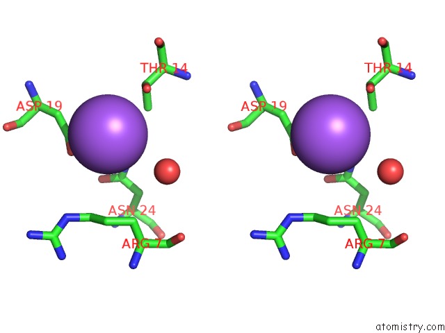
Sodium binding site 1 out of 3 in 6g7o
Go back to
Sodium binding site 1 out
of 3 in the Crystal Structure of Human Alkaline Ceramidase 3 (ACER3) at 2.7 Angstrom Resolution
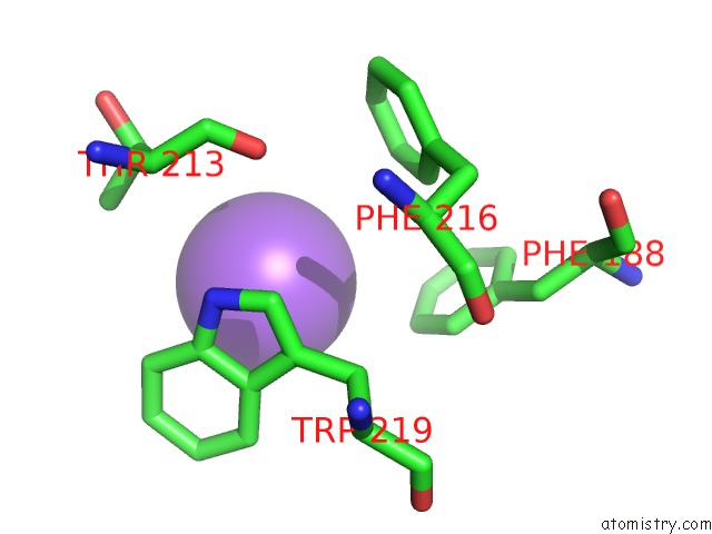
Mono view
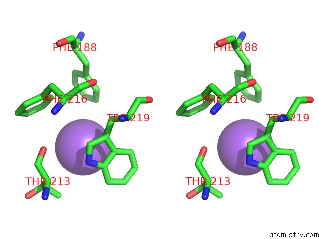
Stereo pair view
Mono view
Stereo pair view
A full contact list of Sodium with other atoms in the Na binding
site number 1 of Crystal Structure of Human Alkaline Ceramidase 3 (ACER3) at 2.7 Angstrom Resolution within 5.0Å range:
|
Sodium binding site 2 out of 3 in 6g7o
Go back to
Sodium binding site 2 out
of 3 in the Crystal Structure of Human Alkaline Ceramidase 3 (ACER3) at 2.7 Angstrom Resolution

Mono view
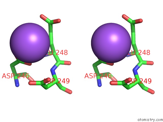
Stereo pair view
Mono view
Stereo pair view
A full contact list of Sodium with other atoms in the Na binding
site number 2 of Crystal Structure of Human Alkaline Ceramidase 3 (ACER3) at 2.7 Angstrom Resolution within 5.0Å range:
|
Sodium binding site 3 out of 3 in 6g7o
Go back to
Sodium binding site 3 out
of 3 in the Crystal Structure of Human Alkaline Ceramidase 3 (ACER3) at 2.7 Angstrom Resolution
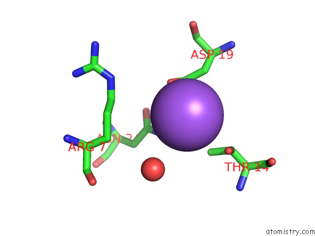
Mono view
Stereo pair view
Mono view
Stereo pair view
A full contact list of Sodium with other atoms in the Na binding
site number 3 of Crystal Structure of Human Alkaline Ceramidase 3 (ACER3) at 2.7 Angstrom Resolution within 5.0Å range:
|
Reference:
I.Vasiliauskaite-Brooks,
R.D.Healey,
P.Rochaix,
J.Saint-Paul,
R.Sounier,
C.Grison,
T.Waltrich-Augusto,
M.Fortier,
F.Hoh,
E.M.Saied,
C.Arenz,
S.Basu,
C.Leyrat,
S.Granier.
Structure of A Human Intramembrane Ceramidase Explains Enzymatic Dysfunction Found in Leukodystrophy. Nat Commun V. 9 5437 2018.
ISSN: ESSN 2041-1723
PubMed: 30575723
DOI: 10.1038/S41467-018-07864-W
Page generated: Tue Oct 8 08:57:52 2024
ISSN: ESSN 2041-1723
PubMed: 30575723
DOI: 10.1038/S41467-018-07864-W
Last articles
Fe in 2YXOFe in 2YRS
Fe in 2YXC
Fe in 2YNM
Fe in 2YVJ
Fe in 2YP1
Fe in 2YU2
Fe in 2YU1
Fe in 2YQB
Fe in 2YOO