Sodium »
PDB 6dq0-6e8t »
6e1r »
Sodium in PDB 6e1r: Crystal Structure of the Acinetobacter Phage VB_APIP_P1 Tailspike Protein
Protein crystallography data
The structure of Crystal Structure of the Acinetobacter Phage VB_APIP_P1 Tailspike Protein, PDB code: 6e1r
was solved by
M.Plattner,
M.M.Shneider,
H.Oliveira,
J.Azeredo,
P.G.Leiman,
with X-Ray Crystallography technique. A brief refinement statistics is given in the table below:
| Resolution Low / High (Å) | 49.12 / 2.69 |
| Space group | P 21 21 21 |
| Cell size a, b, c (Å), α, β, γ (°) | 81.123, 90.015, 508.604, 90.00, 90.00, 90.00 |
| R / Rfree (%) | 20 / 23.8 |
Other elements in 6e1r:
The structure of Crystal Structure of the Acinetobacter Phage VB_APIP_P1 Tailspike Protein also contains other interesting chemical elements:
| Chlorine | (Cl) | 3 atoms |
Sodium Binding Sites:
The binding sites of Sodium atom in the Crystal Structure of the Acinetobacter Phage VB_APIP_P1 Tailspike Protein
(pdb code 6e1r). This binding sites where shown within
5.0 Angstroms radius around Sodium atom.
In total 3 binding sites of Sodium where determined in the Crystal Structure of the Acinetobacter Phage VB_APIP_P1 Tailspike Protein, PDB code: 6e1r:
Jump to Sodium binding site number: 1; 2; 3;
In total 3 binding sites of Sodium where determined in the Crystal Structure of the Acinetobacter Phage VB_APIP_P1 Tailspike Protein, PDB code: 6e1r:
Jump to Sodium binding site number: 1; 2; 3;
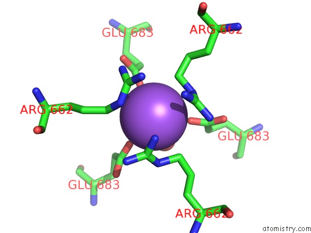
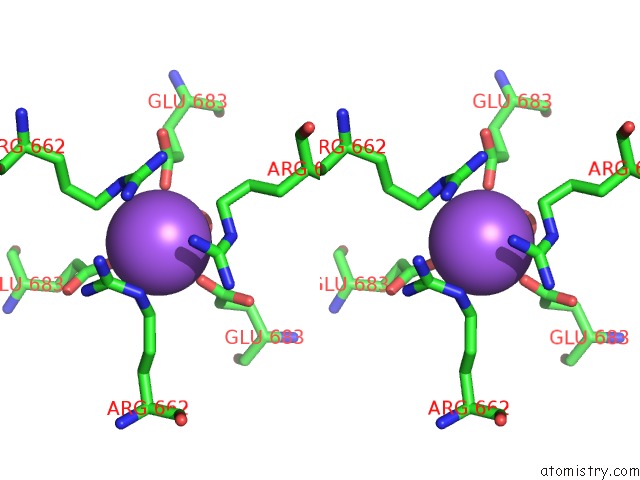
Sodium binding site 1 out of 3 in 6e1r
Go back to
Sodium binding site 1 out
of 3 in the Crystal Structure of the Acinetobacter Phage VB_APIP_P1 Tailspike Protein
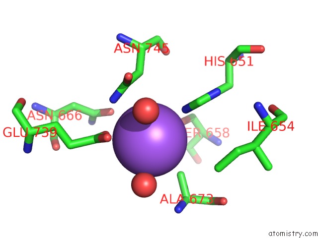
Mono view
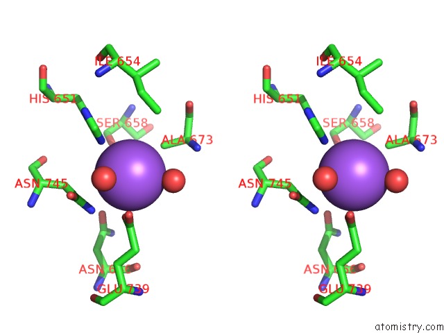
Stereo pair view
Mono view
Stereo pair view
A full contact list of Sodium with other atoms in the Na binding
site number 1 of Crystal Structure of the Acinetobacter Phage VB_APIP_P1 Tailspike Protein within 5.0Å range:
|
Sodium binding site 2 out of 3 in 6e1r
Go back to
Sodium binding site 2 out
of 3 in the Crystal Structure of the Acinetobacter Phage VB_APIP_P1 Tailspike Protein
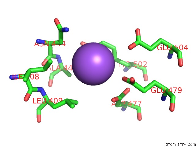
Mono view
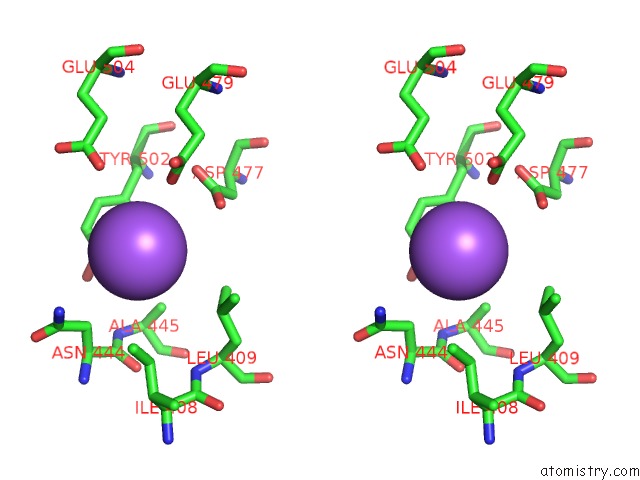
Stereo pair view
Mono view
Stereo pair view
A full contact list of Sodium with other atoms in the Na binding
site number 2 of Crystal Structure of the Acinetobacter Phage VB_APIP_P1 Tailspike Protein within 5.0Å range:
|
Sodium binding site 3 out of 3 in 6e1r
Go back to
Sodium binding site 3 out
of 3 in the Crystal Structure of the Acinetobacter Phage VB_APIP_P1 Tailspike Protein
Mono view
Stereo pair view
Mono view
Stereo pair view
A full contact list of Sodium with other atoms in the Na binding
site number 3 of Crystal Structure of the Acinetobacter Phage VB_APIP_P1 Tailspike Protein within 5.0Å range:
|
Reference:
M.Plattner,
M.M.Shneider,
H.Oliveira,
J.Azeredo,
P.G.Leiman.
Crystal Structure of the Acinetobacter Phage VB_APIP_P1 Tailspike Protein To Be Published.
Page generated: Tue Oct 8 08:14:37 2024
Last articles
Fe in 2YXOFe in 2YRS
Fe in 2YXC
Fe in 2YNM
Fe in 2YVJ
Fe in 2YP1
Fe in 2YU2
Fe in 2YU1
Fe in 2YQB
Fe in 2YOO