Sodium »
PDB 5zxe-6ah7 »
6a0s »
Sodium in PDB 6a0s: Homoserine Dehydrogenase From Thermus Thermophilus HB8 Complexed with Hse and Nadph
Enzymatic activity of Homoserine Dehydrogenase From Thermus Thermophilus HB8 Complexed with Hse and Nadph
All present enzymatic activity of Homoserine Dehydrogenase From Thermus Thermophilus HB8 Complexed with Hse and Nadph:
1.1.1.3;
1.1.1.3;
Protein crystallography data
The structure of Homoserine Dehydrogenase From Thermus Thermophilus HB8 Complexed with Hse and Nadph, PDB code: 6a0s
was solved by
S.Akai,
H.Ikushiro,
T.Sawai,
T.Yano,
N.Kamiya,
I.Miyahara,
with X-Ray Crystallography technique. A brief refinement statistics is given in the table below:
| Resolution Low / High (Å) | 50.00 / 2.00 |
| Space group | P 32 2 1 |
| Cell size a, b, c (Å), α, β, γ (°) | 119.619, 119.619, 144.338, 90.00, 90.00, 120.00 |
| R / Rfree (%) | 15.9 / 20 |
Sodium Binding Sites:
The binding sites of Sodium atom in the Homoserine Dehydrogenase From Thermus Thermophilus HB8 Complexed with Hse and Nadph
(pdb code 6a0s). This binding sites where shown within
5.0 Angstroms radius around Sodium atom.
In total 2 binding sites of Sodium where determined in the Homoserine Dehydrogenase From Thermus Thermophilus HB8 Complexed with Hse and Nadph, PDB code: 6a0s:
Jump to Sodium binding site number: 1; 2;
In total 2 binding sites of Sodium where determined in the Homoserine Dehydrogenase From Thermus Thermophilus HB8 Complexed with Hse and Nadph, PDB code: 6a0s:
Jump to Sodium binding site number: 1; 2;
Sodium binding site 1 out of 2 in 6a0s
Go back to
Sodium binding site 1 out
of 2 in the Homoserine Dehydrogenase From Thermus Thermophilus HB8 Complexed with Hse and Nadph
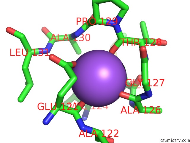
Mono view
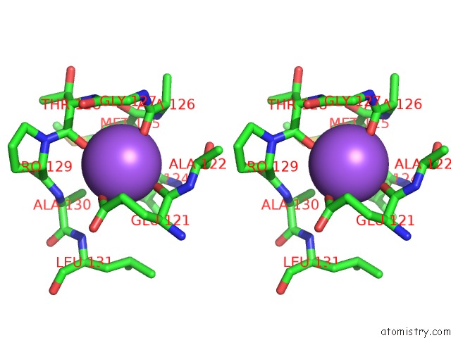
Stereo pair view
Mono view
Stereo pair view
A full contact list of Sodium with other atoms in the Na binding
site number 1 of Homoserine Dehydrogenase From Thermus Thermophilus HB8 Complexed with Hse and Nadph within 5.0Å range:
|
Sodium binding site 2 out of 2 in 6a0s
Go back to
Sodium binding site 2 out
of 2 in the Homoserine Dehydrogenase From Thermus Thermophilus HB8 Complexed with Hse and Nadph
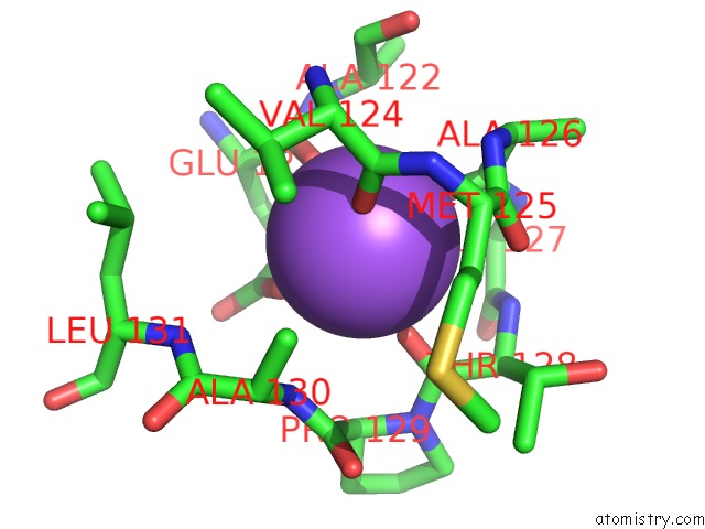
Mono view
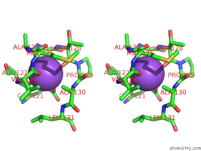
Stereo pair view
Mono view
Stereo pair view
A full contact list of Sodium with other atoms in the Na binding
site number 2 of Homoserine Dehydrogenase From Thermus Thermophilus HB8 Complexed with Hse and Nadph within 5.0Å range:
|
Reference:
S.Akai,
H.Ikushiro,
T.Sawai,
T.Yano,
N.Kamiya,
I.Miyahara.
The Crystal Structure of Homoserine Dehydrogenase Complexed with L-Homoserine and Nadph in A Closed Form J. Biochem. V. 165 185 2019.
ISSN: ISSN 1756-2651
PubMed: 30423116
DOI: 10.1093/JB/MVY094
Page generated: Mon Aug 18 03:30:51 2025
ISSN: ISSN 1756-2651
PubMed: 30423116
DOI: 10.1093/JB/MVY094
Last articles
Na in 7RLRNa in 7RJR
Na in 7RJ9
Na in 7RJO
Na in 7RJI
Na in 7RG8
Na in 7RIZ
Na in 7RCP
Na in 7RIS
Na in 7RHS