Sodium »
PDB 5xvd-5ysu »
5yid »
Sodium in PDB 5yid: Crystal Structure of Kni-10395 Bound Plasmepsin II (Pmii) From Plasmodium Falciparum
Enzymatic activity of Crystal Structure of Kni-10395 Bound Plasmepsin II (Pmii) From Plasmodium Falciparum
All present enzymatic activity of Crystal Structure of Kni-10395 Bound Plasmepsin II (Pmii) From Plasmodium Falciparum:
3.4.23.39;
3.4.23.39;
Protein crystallography data
The structure of Crystal Structure of Kni-10395 Bound Plasmepsin II (Pmii) From Plasmodium Falciparum, PDB code: 5yid
was solved by
V.Mishra,
I.Rathore,
P.Bhaumik,
with X-Ray Crystallography technique. A brief refinement statistics is given in the table below:
| Resolution Low / High (Å) | 38.00 / 2.10 |
| Space group | I 4 |
| Cell size a, b, c (Å), α, β, γ (°) | 107.600, 107.600, 72.560, 90.00, 90.00, 90.00 |
| R / Rfree (%) | 18.8 / 22.5 |
Sodium Binding Sites:
The binding sites of Sodium atom in the Crystal Structure of Kni-10395 Bound Plasmepsin II (Pmii) From Plasmodium Falciparum
(pdb code 5yid). This binding sites where shown within
5.0 Angstroms radius around Sodium atom.
In total only one binding site of Sodium was determined in the Crystal Structure of Kni-10395 Bound Plasmepsin II (Pmii) From Plasmodium Falciparum, PDB code: 5yid:
In total only one binding site of Sodium was determined in the Crystal Structure of Kni-10395 Bound Plasmepsin II (Pmii) From Plasmodium Falciparum, PDB code: 5yid:
Sodium binding site 1 out of 1 in 5yid
Go back to
Sodium binding site 1 out
of 1 in the Crystal Structure of Kni-10395 Bound Plasmepsin II (Pmii) From Plasmodium Falciparum
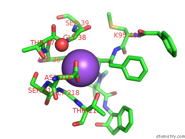
Mono view
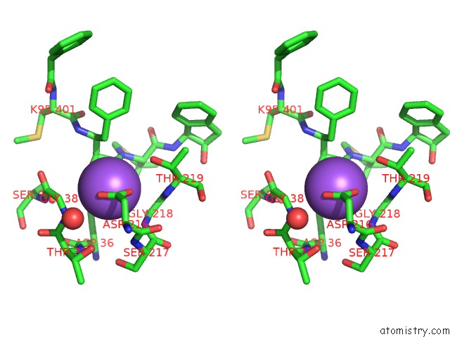
Stereo pair view
Mono view
Stereo pair view
A full contact list of Sodium with other atoms in the Na binding
site number 1 of Crystal Structure of Kni-10395 Bound Plasmepsin II (Pmii) From Plasmodium Falciparum within 5.0Å range:
|
Reference:
V.Mishra,
I.Rathore,
A.Arekar,
L.K.Sthanam,
H.Xiao,
Y.Kiso,
S.Sen,
S.Patankar,
A.Gustchina,
K.Hidaka,
A.Wlodawer,
R.Y.Yada,
P.Bhaumik.
Deciphering the Mechanism of Potent Peptidomimetic Inhibitors Targeting Plasmepsins - Biochemical and Structural Insights. Febs J. V. 285 3077 2018.
ISSN: ISSN 1742-464X
PubMed: 29943906
DOI: 10.1111/FEBS.14598
Page generated: Tue Oct 8 01:23:19 2024
ISSN: ISSN 1742-464X
PubMed: 29943906
DOI: 10.1111/FEBS.14598
Last articles
Zn in 9J0NZn in 9J0O
Zn in 9J0P
Zn in 9FJX
Zn in 9EKB
Zn in 9C0F
Zn in 9CAH
Zn in 9CH0
Zn in 9CH3
Zn in 9CH1