Sodium »
PDB 5v0e-5vb8 »
5v9g »
Sodium in PDB 5v9g: Structure of the H477R Variant of Rat Cytosolic Pepck in Complex with Oxalate and Gtp.
Enzymatic activity of Structure of the H477R Variant of Rat Cytosolic Pepck in Complex with Oxalate and Gtp.
All present enzymatic activity of Structure of the H477R Variant of Rat Cytosolic Pepck in Complex with Oxalate and Gtp.:
4.1.1.32;
4.1.1.32;
Protein crystallography data
The structure of Structure of the H477R Variant of Rat Cytosolic Pepck in Complex with Oxalate and Gtp., PDB code: 5v9g
was solved by
T.Holyoak,
D.S.Cui,
with X-Ray Crystallography technique. A brief refinement statistics is given in the table below:
| Resolution Low / High (Å) | 28.37 / 1.95 |
| Space group | P 1 21 1 |
| Cell size a, b, c (Å), α, β, γ (°) | 46.526, 119.605, 60.679, 90.00, 106.83, 90.00 |
| R / Rfree (%) | 18.4 / 21.2 |
Other elements in 5v9g:
The structure of Structure of the H477R Variant of Rat Cytosolic Pepck in Complex with Oxalate and Gtp. also contains other interesting chemical elements:
| Manganese | (Mn) | 2 atoms |
Sodium Binding Sites:
The binding sites of Sodium atom in the Structure of the H477R Variant of Rat Cytosolic Pepck in Complex with Oxalate and Gtp.
(pdb code 5v9g). This binding sites where shown within
5.0 Angstroms radius around Sodium atom.
In total only one binding site of Sodium was determined in the Structure of the H477R Variant of Rat Cytosolic Pepck in Complex with Oxalate and Gtp., PDB code: 5v9g:
In total only one binding site of Sodium was determined in the Structure of the H477R Variant of Rat Cytosolic Pepck in Complex with Oxalate and Gtp., PDB code: 5v9g:
Sodium binding site 1 out of 1 in 5v9g
Go back to
Sodium binding site 1 out
of 1 in the Structure of the H477R Variant of Rat Cytosolic Pepck in Complex with Oxalate and Gtp.
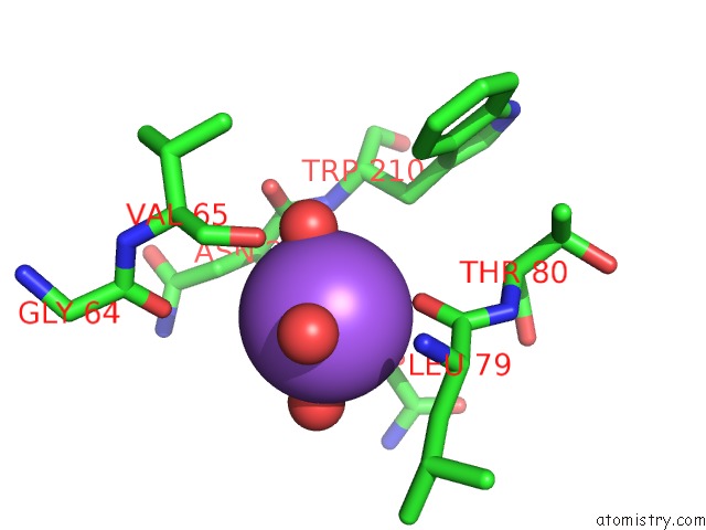
Mono view
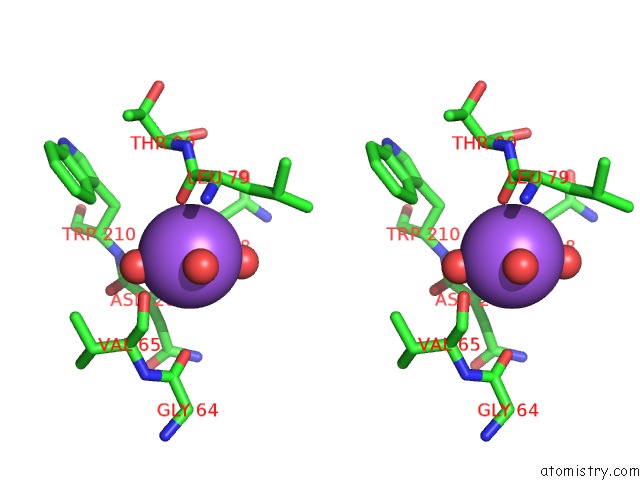
Stereo pair view
Mono view
Stereo pair view
A full contact list of Sodium with other atoms in the Na binding
site number 1 of Structure of the H477R Variant of Rat Cytosolic Pepck in Complex with Oxalate and Gtp. within 5.0Å range:
|
Reference:
D.S.Cui,
A.Broom,
M.J.Mcleod,
E.M.Meiering,
T.Holyoak.
Asymmetric Anchoring Is Required For Efficient Omega-Loop Opening and Closing in Cytosolic Phosphoenolpyruvate Carboxykinase. Biochemistry V. 56 2106 2017.
ISSN: ISSN 1520-4995
PubMed: 28345895
DOI: 10.1021/ACS.BIOCHEM.7B00178
Page generated: Tue Oct 8 00:46:50 2024
ISSN: ISSN 1520-4995
PubMed: 28345895
DOI: 10.1021/ACS.BIOCHEM.7B00178
Last articles
Zn in 9J0NZn in 9J0O
Zn in 9J0P
Zn in 9FJX
Zn in 9EKB
Zn in 9C0F
Zn in 9CAH
Zn in 9CH0
Zn in 9CH3
Zn in 9CH1