Sodium »
PDB 5uka-5v0d »
5ur0 »
Sodium in PDB 5ur0: Crystallographic Structure of Glyceraldehyde-3-Phosphate Dehydrogenase From Naegleria Gruberi
Enzymatic activity of Crystallographic Structure of Glyceraldehyde-3-Phosphate Dehydrogenase From Naegleria Gruberi
All present enzymatic activity of Crystallographic Structure of Glyceraldehyde-3-Phosphate Dehydrogenase From Naegleria Gruberi:
1.2.1.12;
1.2.1.12;
Protein crystallography data
The structure of Crystallographic Structure of Glyceraldehyde-3-Phosphate Dehydrogenase From Naegleria Gruberi, PDB code: 5ur0
was solved by
A.T.P.Machado,
M.Silva,
J.Iulek,
with X-Ray Crystallography technique. A brief refinement statistics is given in the table below:
| Resolution Low / High (Å) | 29.85 / 1.94 |
| Space group | P 1 21 1 |
| Cell size a, b, c (Å), α, β, γ (°) | 83.743, 94.552, 90.932, 90.00, 99.96, 90.00 |
| R / Rfree (%) | 15.4 / 19.8 |
Sodium Binding Sites:
The binding sites of Sodium atom in the Crystallographic Structure of Glyceraldehyde-3-Phosphate Dehydrogenase From Naegleria Gruberi
(pdb code 5ur0). This binding sites where shown within
5.0 Angstroms radius around Sodium atom.
In total 4 binding sites of Sodium where determined in the Crystallographic Structure of Glyceraldehyde-3-Phosphate Dehydrogenase From Naegleria Gruberi, PDB code: 5ur0:
Jump to Sodium binding site number: 1; 2; 3; 4;
In total 4 binding sites of Sodium where determined in the Crystallographic Structure of Glyceraldehyde-3-Phosphate Dehydrogenase From Naegleria Gruberi, PDB code: 5ur0:
Jump to Sodium binding site number: 1; 2; 3; 4;
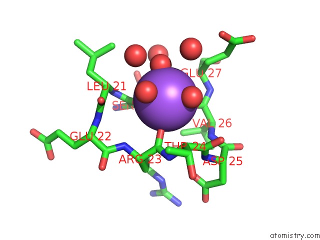
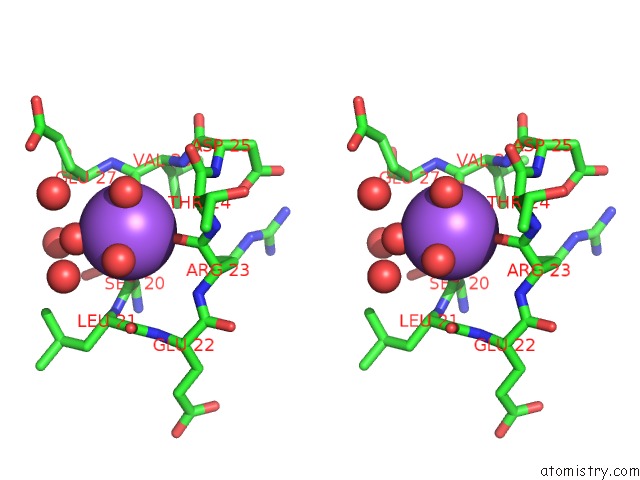
Sodium binding site 1 out of 4 in 5ur0
Go back to
Sodium binding site 1 out
of 4 in the Crystallographic Structure of Glyceraldehyde-3-Phosphate Dehydrogenase From Naegleria Gruberi
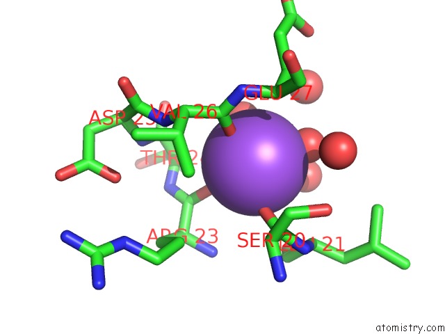
Mono view
Stereo pair view
Mono view
Stereo pair view
A full contact list of Sodium with other atoms in the Na binding
site number 1 of Crystallographic Structure of Glyceraldehyde-3-Phosphate Dehydrogenase From Naegleria Gruberi within 5.0Å range:
|
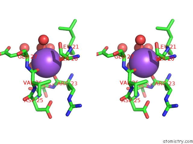
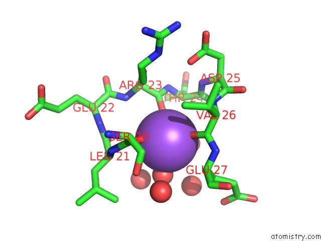
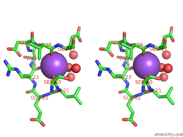
Sodium binding site 2 out of 4 in 5ur0
Go back to
Sodium binding site 2 out
of 4 in the Crystallographic Structure of Glyceraldehyde-3-Phosphate Dehydrogenase From Naegleria Gruberi
Mono view
Stereo pair view
Mono view
Stereo pair view
A full contact list of Sodium with other atoms in the Na binding
site number 2 of Crystallographic Structure of Glyceraldehyde-3-Phosphate Dehydrogenase From Naegleria Gruberi within 5.0Å range:
|
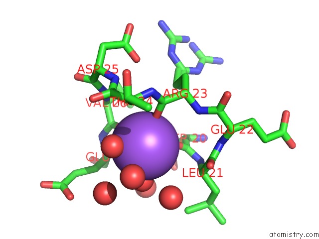
Sodium binding site 3 out of 4 in 5ur0
Go back to
Sodium binding site 3 out
of 4 in the Crystallographic Structure of Glyceraldehyde-3-Phosphate Dehydrogenase From Naegleria Gruberi
Mono view
Stereo pair view
Mono view
Stereo pair view
A full contact list of Sodium with other atoms in the Na binding
site number 3 of Crystallographic Structure of Glyceraldehyde-3-Phosphate Dehydrogenase From Naegleria Gruberi within 5.0Å range:
|
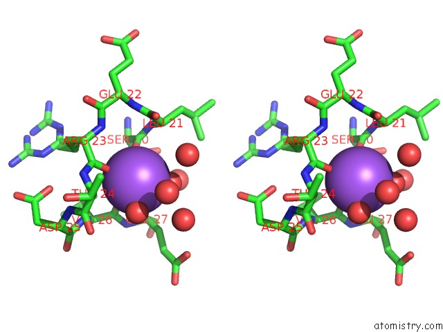
Sodium binding site 4 out of 4 in 5ur0
Go back to
Sodium binding site 4 out
of 4 in the Crystallographic Structure of Glyceraldehyde-3-Phosphate Dehydrogenase From Naegleria Gruberi
Mono view
Stereo pair view
Mono view
Stereo pair view
A full contact list of Sodium with other atoms in the Na binding
site number 4 of Crystallographic Structure of Glyceraldehyde-3-Phosphate Dehydrogenase From Naegleria Gruberi within 5.0Å range:
|
Reference:
A.T.P.Machado,
M.Silva,
J.Iulek.
Structural Studies of Glyceraldehyde-3-Phosphate Dehydrogenase From Naegleria Gruberi, the First One From Phylum Percolozoa. Biochim. Biophys. Acta V.1866 581 2018.
ISSN: ISSN 0006-3002
PubMed: 29501559
DOI: 10.1016/J.BBAPAP.2018.02.006
Page generated: Tue Oct 8 00:38:58 2024
ISSN: ISSN 0006-3002
PubMed: 29501559
DOI: 10.1016/J.BBAPAP.2018.02.006
Last articles
Zn in 9J0NZn in 9J0O
Zn in 9J0P
Zn in 9FJX
Zn in 9EKB
Zn in 9C0F
Zn in 9CAH
Zn in 9CH0
Zn in 9CH3
Zn in 9CH1