Sodium »
PDB 5txu-5u63 »
5tyd »
Sodium in PDB 5tyd: Dna Polymerase Mu Reactant Complex, 10 Mm MG2+ (45 Min)
Enzymatic activity of Dna Polymerase Mu Reactant Complex, 10 Mm MG2+ (45 Min)
All present enzymatic activity of Dna Polymerase Mu Reactant Complex, 10 Mm MG2+ (45 Min):
2.7.7.7;
2.7.7.7;
Protein crystallography data
The structure of Dna Polymerase Mu Reactant Complex, 10 Mm MG2+ (45 Min), PDB code: 5tyd
was solved by
J.A.Jamsen,
S.H.Wilson,
with X-Ray Crystallography technique. A brief refinement statistics is given in the table below:
| Resolution Low / High (Å) | 34.94 / 1.90 |
| Space group | P 21 21 21 |
| Cell size a, b, c (Å), α, β, γ (°) | 59.891, 68.590, 110.476, 90.00, 90.00, 90.00 |
| R / Rfree (%) | 17.1 / 21.2 |
Other elements in 5tyd:
The structure of Dna Polymerase Mu Reactant Complex, 10 Mm MG2+ (45 Min) also contains other interesting chemical elements:
| Magnesium | (Mg) | 2 atoms |
Sodium Binding Sites:
The binding sites of Sodium atom in the Dna Polymerase Mu Reactant Complex, 10 Mm MG2+ (45 Min)
(pdb code 5tyd). This binding sites where shown within
5.0 Angstroms radius around Sodium atom.
In total only one binding site of Sodium was determined in the Dna Polymerase Mu Reactant Complex, 10 Mm MG2+ (45 Min), PDB code: 5tyd:
In total only one binding site of Sodium was determined in the Dna Polymerase Mu Reactant Complex, 10 Mm MG2+ (45 Min), PDB code: 5tyd:
Sodium binding site 1 out of 1 in 5tyd
Go back to
Sodium binding site 1 out
of 1 in the Dna Polymerase Mu Reactant Complex, 10 Mm MG2+ (45 Min)
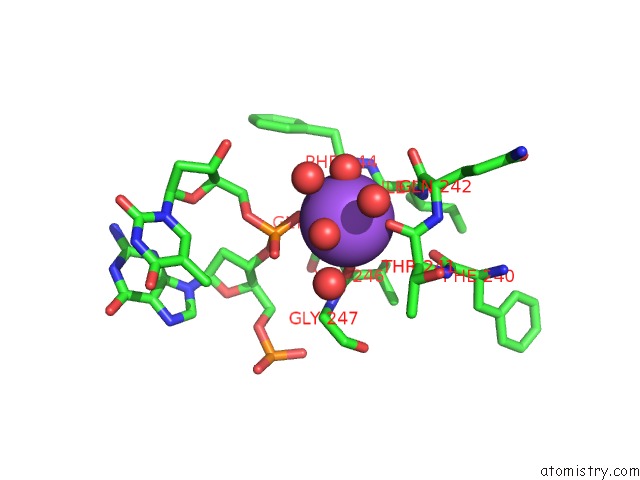
Mono view
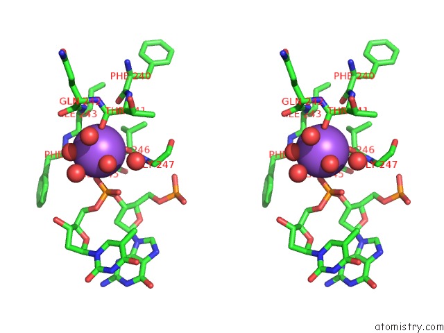
Stereo pair view
Mono view
Stereo pair view
A full contact list of Sodium with other atoms in the Na binding
site number 1 of Dna Polymerase Mu Reactant Complex, 10 Mm MG2+ (45 Min) within 5.0Å range:
|
Reference:
J.A.Jamsen,
W.A.Beard,
L.C.Pedersen,
D.D.Shock,
A.F.Moon,
J.M.Krahn,
K.Bebenek,
T.A.Kunkel,
S.H.Wilson.
Time-Lapse Crystallography Snapshots of A Double-Strand Break Repair Polymerase in Action. Nat Commun V. 8 253 2017.
ISSN: ESSN 2041-1723
PubMed: 28811466
DOI: 10.1038/S41467-017-00271-7
Page generated: Tue Oct 8 00:18:46 2024
ISSN: ESSN 2041-1723
PubMed: 28811466
DOI: 10.1038/S41467-017-00271-7
Last articles
Zn in 9MJ5Zn in 9HNW
Zn in 9G0L
Zn in 9FNE
Zn in 9DZN
Zn in 9E0I
Zn in 9D32
Zn in 9DAK
Zn in 8ZXC
Zn in 8ZUF