Sodium »
PDB 5syu-5tde »
5tcd »
Sodium in PDB 5tcd: Human Alkaline Sphingomyelinase (ENPP7) in Complex with Phosphocholine
Enzymatic activity of Human Alkaline Sphingomyelinase (ENPP7) in Complex with Phosphocholine
All present enzymatic activity of Human Alkaline Sphingomyelinase (ENPP7) in Complex with Phosphocholine:
3.1.4.12;
3.1.4.12;
Protein crystallography data
The structure of Human Alkaline Sphingomyelinase (ENPP7) in Complex with Phosphocholine, PDB code: 5tcd
was solved by
A.Gorelik,
F.Liu,
K.Illes,
B.Nagar,
with X-Ray Crystallography technique. A brief refinement statistics is given in the table below:
| Resolution Low / High (Å) | 38.44 / 2.40 |
| Space group | P 31 2 1 |
| Cell size a, b, c (Å), α, β, γ (°) | 104.223, 104.223, 113.893, 90.00, 90.00, 120.00 |
| R / Rfree (%) | 17.3 / 20.8 |
Other elements in 5tcd:
The structure of Human Alkaline Sphingomyelinase (ENPP7) in Complex with Phosphocholine also contains other interesting chemical elements:
| Zinc | (Zn) | 2 atoms |
| Iodine | (I) | 37 atoms |
Sodium Binding Sites:
The binding sites of Sodium atom in the Human Alkaline Sphingomyelinase (ENPP7) in Complex with Phosphocholine
(pdb code 5tcd). This binding sites where shown within
5.0 Angstroms radius around Sodium atom.
In total only one binding site of Sodium was determined in the Human Alkaline Sphingomyelinase (ENPP7) in Complex with Phosphocholine, PDB code: 5tcd:
In total only one binding site of Sodium was determined in the Human Alkaline Sphingomyelinase (ENPP7) in Complex with Phosphocholine, PDB code: 5tcd:
Sodium binding site 1 out of 1 in 5tcd
Go back to
Sodium binding site 1 out
of 1 in the Human Alkaline Sphingomyelinase (ENPP7) in Complex with Phosphocholine
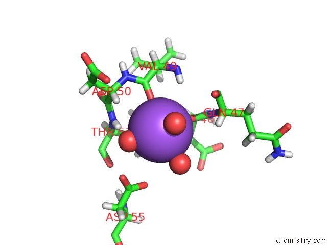
Mono view
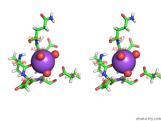
Stereo pair view
Mono view
Stereo pair view
A full contact list of Sodium with other atoms in the Na binding
site number 1 of Human Alkaline Sphingomyelinase (ENPP7) in Complex with Phosphocholine within 5.0Å range:
|
Reference:
A.Gorelik,
F.Liu,
K.Illes,
B.Nagar.
Crystal Structure of the Human Alkaline Sphingomyelinase Provides Insights Into Substrate Recognition. J. Biol. Chem. V. 292 7087 2017.
ISSN: ESSN 1083-351X
PubMed: 28292932
DOI: 10.1074/JBC.M116.769273
Page generated: Tue Oct 8 00:07:44 2024
ISSN: ESSN 1083-351X
PubMed: 28292932
DOI: 10.1074/JBC.M116.769273
Last articles
Zn in 9MJ5Zn in 9HNW
Zn in 9G0L
Zn in 9FNE
Zn in 9DZN
Zn in 9E0I
Zn in 9D32
Zn in 9DAK
Zn in 8ZXC
Zn in 8ZUF