Sodium »
PDB 5syu-5tde »
5t25 »
Sodium in PDB 5t25: Kinetic, Spectral and Structural Characterization of the Slow Binding Inhibitor Acetopyruvate with Dihydrodipicolinate Synthase From Escherichia Coli.
Enzymatic activity of Kinetic, Spectral and Structural Characterization of the Slow Binding Inhibitor Acetopyruvate with Dihydrodipicolinate Synthase From Escherichia Coli.
All present enzymatic activity of Kinetic, Spectral and Structural Characterization of the Slow Binding Inhibitor Acetopyruvate with Dihydrodipicolinate Synthase From Escherichia Coli.:
4.3.3.7;
4.3.3.7;
Protein crystallography data
The structure of Kinetic, Spectral and Structural Characterization of the Slow Binding Inhibitor Acetopyruvate with Dihydrodipicolinate Synthase From Escherichia Coli., PDB code: 5t25
was solved by
L.Chooback,
L.M.Thomas,
W.E.Karsten,
C.D.Fleming,
P.Seabourn,
with X-Ray Crystallography technique. A brief refinement statistics is given in the table below:
| Resolution Low / High (Å) | 26.81 / 1.99 |
| Space group | C 1 2 1 |
| Cell size a, b, c (Å), α, β, γ (°) | 136.647, 56.385, 101.530, 90.00, 127.62, 90.00 |
| R / Rfree (%) | 15.8 / 21.1 |
Sodium Binding Sites:
The binding sites of Sodium atom in the Kinetic, Spectral and Structural Characterization of the Slow Binding Inhibitor Acetopyruvate with Dihydrodipicolinate Synthase From Escherichia Coli.
(pdb code 5t25). This binding sites where shown within
5.0 Angstroms radius around Sodium atom.
In total 10 binding sites of Sodium where determined in the Kinetic, Spectral and Structural Characterization of the Slow Binding Inhibitor Acetopyruvate with Dihydrodipicolinate Synthase From Escherichia Coli., PDB code: 5t25:
Jump to Sodium binding site number: 1; 2; 3; 4; 5; 6; 7; 8; 9; 10;
In total 10 binding sites of Sodium where determined in the Kinetic, Spectral and Structural Characterization of the Slow Binding Inhibitor Acetopyruvate with Dihydrodipicolinate Synthase From Escherichia Coli., PDB code: 5t25:
Jump to Sodium binding site number: 1; 2; 3; 4; 5; 6; 7; 8; 9; 10;
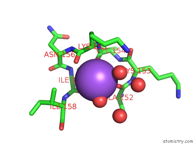
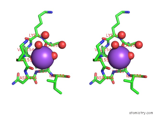
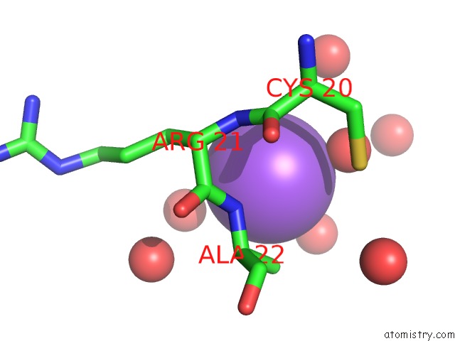
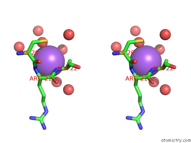
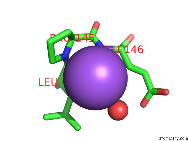
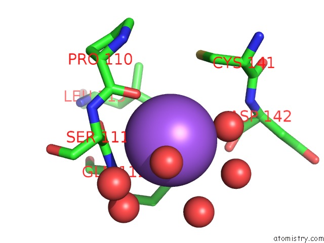
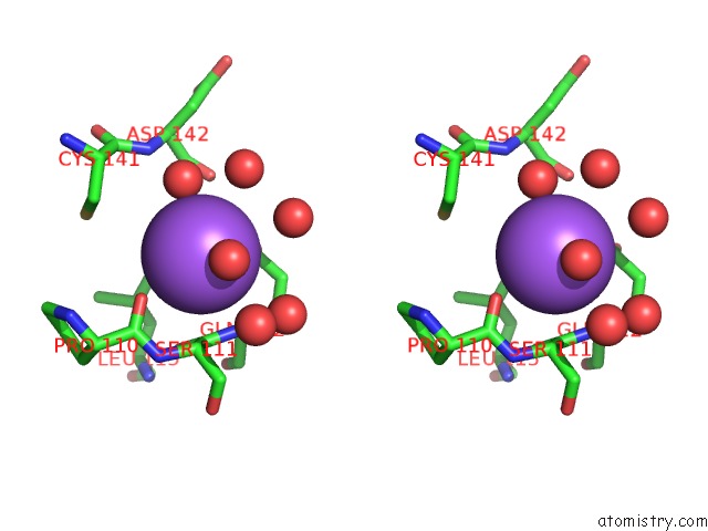
Sodium binding site 1 out of 10 in 5t25
Go back to
Sodium binding site 1 out
of 10 in the Kinetic, Spectral and Structural Characterization of the Slow Binding Inhibitor Acetopyruvate with Dihydrodipicolinate Synthase From Escherichia Coli.
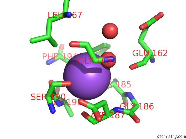
Mono view
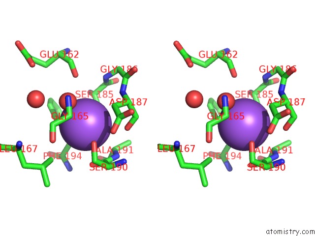
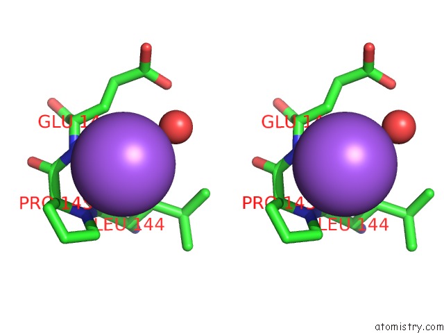
Stereo pair view
Mono view
Stereo pair view
A full contact list of Sodium with other atoms in the Na binding
site number 1 of Kinetic, Spectral and Structural Characterization of the Slow Binding Inhibitor Acetopyruvate with Dihydrodipicolinate Synthase From Escherichia Coli. within 5.0Å range:
|
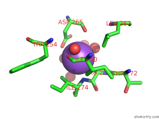
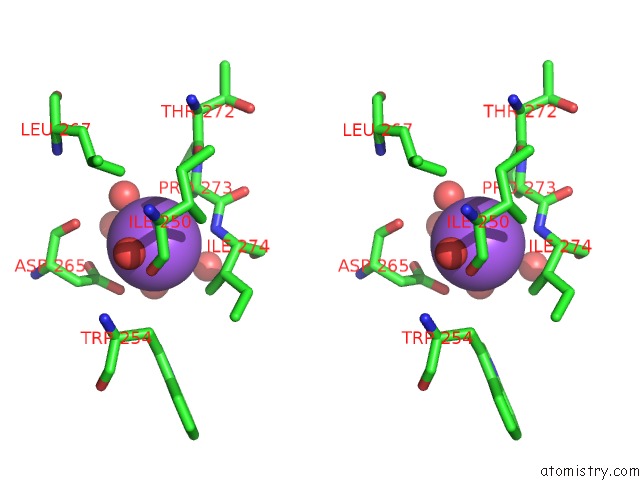
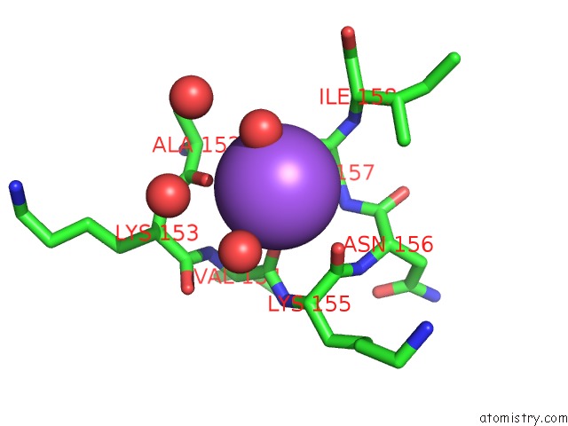
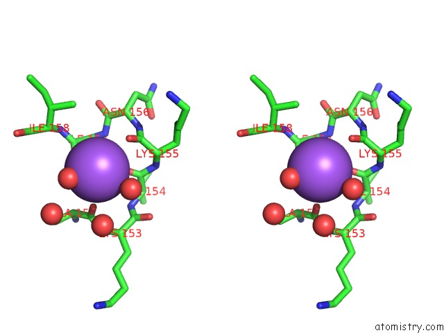
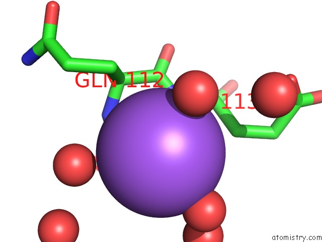
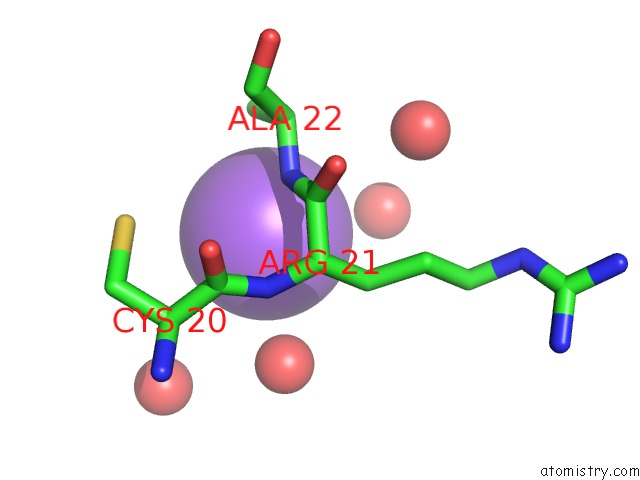
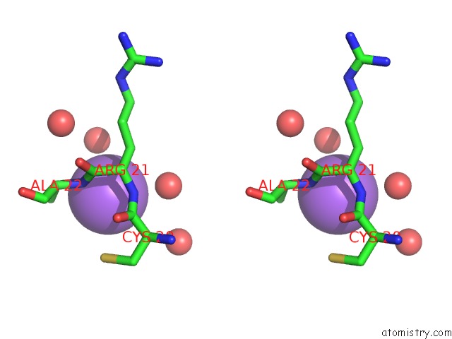
Sodium binding site 2 out of 10 in 5t25
Go back to
Sodium binding site 2 out
of 10 in the Kinetic, Spectral and Structural Characterization of the Slow Binding Inhibitor Acetopyruvate with Dihydrodipicolinate Synthase From Escherichia Coli.
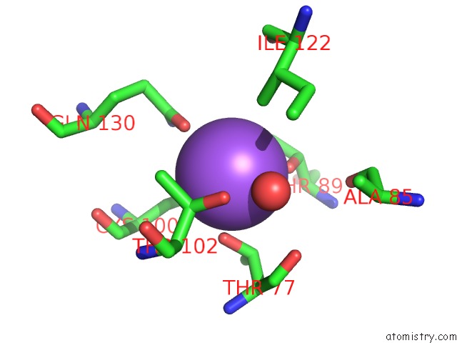
Mono view
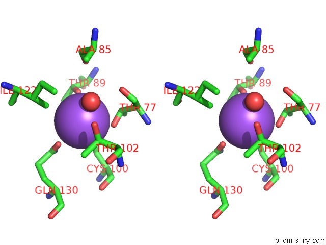
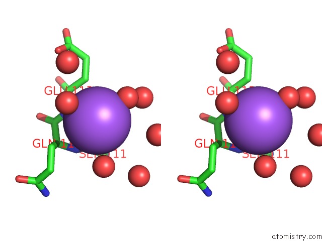
Stereo pair view
Mono view
Stereo pair view
A full contact list of Sodium with other atoms in the Na binding
site number 2 of Kinetic, Spectral and Structural Characterization of the Slow Binding Inhibitor Acetopyruvate with Dihydrodipicolinate Synthase From Escherichia Coli. within 5.0Å range:
|
Sodium binding site 3 out of 10 in 5t25
Go back to
Sodium binding site 3 out
of 10 in the Kinetic, Spectral and Structural Characterization of the Slow Binding Inhibitor Acetopyruvate with Dihydrodipicolinate Synthase From Escherichia Coli.
Mono view
Stereo pair view
Mono view
Stereo pair view
A full contact list of Sodium with other atoms in the Na binding
site number 3 of Kinetic, Spectral and Structural Characterization of the Slow Binding Inhibitor Acetopyruvate with Dihydrodipicolinate Synthase From Escherichia Coli. within 5.0Å range:
|
Sodium binding site 4 out of 10 in 5t25
Go back to
Sodium binding site 4 out
of 10 in the Kinetic, Spectral and Structural Characterization of the Slow Binding Inhibitor Acetopyruvate with Dihydrodipicolinate Synthase From Escherichia Coli.
Mono view
Stereo pair view
Mono view
Stereo pair view
A full contact list of Sodium with other atoms in the Na binding
site number 4 of Kinetic, Spectral and Structural Characterization of the Slow Binding Inhibitor Acetopyruvate with Dihydrodipicolinate Synthase From Escherichia Coli. within 5.0Å range:
|
Sodium binding site 5 out of 10 in 5t25
Go back to
Sodium binding site 5 out
of 10 in the Kinetic, Spectral and Structural Characterization of the Slow Binding Inhibitor Acetopyruvate with Dihydrodipicolinate Synthase From Escherichia Coli.
Mono view
Stereo pair view
Mono view
Stereo pair view
A full contact list of Sodium with other atoms in the Na binding
site number 5 of Kinetic, Spectral and Structural Characterization of the Slow Binding Inhibitor Acetopyruvate with Dihydrodipicolinate Synthase From Escherichia Coli. within 5.0Å range:
|
Sodium binding site 6 out of 10 in 5t25
Go back to
Sodium binding site 6 out
of 10 in the Kinetic, Spectral and Structural Characterization of the Slow Binding Inhibitor Acetopyruvate with Dihydrodipicolinate Synthase From Escherichia Coli.
Mono view
Stereo pair view
Mono view
Stereo pair view
A full contact list of Sodium with other atoms in the Na binding
site number 6 of Kinetic, Spectral and Structural Characterization of the Slow Binding Inhibitor Acetopyruvate with Dihydrodipicolinate Synthase From Escherichia Coli. within 5.0Å range:
|
Sodium binding site 7 out of 10 in 5t25
Go back to
Sodium binding site 7 out
of 10 in the Kinetic, Spectral and Structural Characterization of the Slow Binding Inhibitor Acetopyruvate with Dihydrodipicolinate Synthase From Escherichia Coli.
Mono view
Stereo pair view
Mono view
Stereo pair view
A full contact list of Sodium with other atoms in the Na binding
site number 7 of Kinetic, Spectral and Structural Characterization of the Slow Binding Inhibitor Acetopyruvate with Dihydrodipicolinate Synthase From Escherichia Coli. within 5.0Å range:
|
Sodium binding site 8 out of 10 in 5t25
Go back to
Sodium binding site 8 out
of 10 in the Kinetic, Spectral and Structural Characterization of the Slow Binding Inhibitor Acetopyruvate with Dihydrodipicolinate Synthase From Escherichia Coli.
Mono view
Stereo pair view
Mono view
Stereo pair view
A full contact list of Sodium with other atoms in the Na binding
site number 8 of Kinetic, Spectral and Structural Characterization of the Slow Binding Inhibitor Acetopyruvate with Dihydrodipicolinate Synthase From Escherichia Coli. within 5.0Å range:
|
Sodium binding site 9 out of 10 in 5t25
Go back to
Sodium binding site 9 out
of 10 in the Kinetic, Spectral and Structural Characterization of the Slow Binding Inhibitor Acetopyruvate with Dihydrodipicolinate Synthase From Escherichia Coli.
Mono view
Stereo pair view
Mono view
Stereo pair view
A full contact list of Sodium with other atoms in the Na binding
site number 9 of Kinetic, Spectral and Structural Characterization of the Slow Binding Inhibitor Acetopyruvate with Dihydrodipicolinate Synthase From Escherichia Coli. within 5.0Å range:
|
Sodium binding site 10 out of 10 in 5t25
Go back to
Sodium binding site 10 out
of 10 in the Kinetic, Spectral and Structural Characterization of the Slow Binding Inhibitor Acetopyruvate with Dihydrodipicolinate Synthase From Escherichia Coli.
Mono view
Stereo pair view
Mono view
Stereo pair view
A full contact list of Sodium with other atoms in the Na binding
site number 10 of Kinetic, Spectral and Structural Characterization of the Slow Binding Inhibitor Acetopyruvate with Dihydrodipicolinate Synthase From Escherichia Coli. within 5.0Å range:
|
Reference:
L.Chooback,
L.M.Thomas,
W.E.Karsten,
C.D.Fleming,
P.Seabourn.
Kinetic, Spectral and Structural Characterization of the Slow Binding Inhibitor Acetopyruvate with Dihydrodipicolinate Synthase From Escherichia Coli. To Be Published.
Page generated: Tue Oct 8 00:00:53 2024
Last articles
Cl in 5STCCl in 5ST4
Cl in 5SSB
Cl in 5SQR
Cl in 5SPQ
Cl in 5SQH
Cl in 5SPO
Cl in 5SPK
Cl in 5SP4
Cl in 5SPJ