Sodium »
PDB 5o8n-5opd »
5ojo »
Sodium in PDB 5ojo: Sirtuin 5 From Danio Rerio in Complex with 3-Hydroxy-3-Methylglutaryl- CPS1 Peptide
Enzymatic activity of Sirtuin 5 From Danio Rerio in Complex with 3-Hydroxy-3-Methylglutaryl- CPS1 Peptide
All present enzymatic activity of Sirtuin 5 From Danio Rerio in Complex with 3-Hydroxy-3-Methylglutaryl- CPS1 Peptide:
6.3.4.16;
6.3.4.16;
Protein crystallography data
The structure of Sirtuin 5 From Danio Rerio in Complex with 3-Hydroxy-3-Methylglutaryl- CPS1 Peptide, PDB code: 5ojo
was solved by
M.Pannek,
C.Steegborn,
with X-Ray Crystallography technique. A brief refinement statistics is given in the table below:
| Resolution Low / High (Å) | 48.67 / 3.10 |
| Space group | P 65 2 2 |
| Cell size a, b, c (Å), α, β, γ (°) | 87.540, 87.540, 316.920, 90.00, 90.00, 120.00 |
| R / Rfree (%) | 19.6 / 26.6 |
Other elements in 5ojo:
The structure of Sirtuin 5 From Danio Rerio in Complex with 3-Hydroxy-3-Methylglutaryl- CPS1 Peptide also contains other interesting chemical elements:
| Zinc | (Zn) | 2 atoms |
Sodium Binding Sites:
The binding sites of Sodium atom in the Sirtuin 5 From Danio Rerio in Complex with 3-Hydroxy-3-Methylglutaryl- CPS1 Peptide
(pdb code 5ojo). This binding sites where shown within
5.0 Angstroms radius around Sodium atom.
In total only one binding site of Sodium was determined in the Sirtuin 5 From Danio Rerio in Complex with 3-Hydroxy-3-Methylglutaryl- CPS1 Peptide, PDB code: 5ojo:
In total only one binding site of Sodium was determined in the Sirtuin 5 From Danio Rerio in Complex with 3-Hydroxy-3-Methylglutaryl- CPS1 Peptide, PDB code: 5ojo:
Sodium binding site 1 out of 1 in 5ojo
Go back to
Sodium binding site 1 out
of 1 in the Sirtuin 5 From Danio Rerio in Complex with 3-Hydroxy-3-Methylglutaryl- CPS1 Peptide
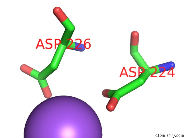
Mono view
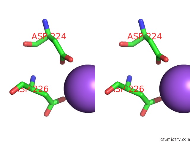
Stereo pair view
Mono view
Stereo pair view
A full contact list of Sodium with other atoms in the Na binding
site number 1 of Sirtuin 5 From Danio Rerio in Complex with 3-Hydroxy-3-Methylglutaryl- CPS1 Peptide within 5.0Å range:
|
Reference:
M.Pannek,
Z.Simic,
M.Fuszard,
M.Meleshin,
D.Rotili,
A.Mai,
M.Schutkowski,
C.Steegborn.
Crystal Structures of the Mitochondrial Deacylase Sirtuin 4 Reveal Isoform-Specific Acyl Recognition and Regulation Features. Nat Commun V. 8 1513 2017.
ISSN: ESSN 2041-1723
PubMed: 29138502
DOI: 10.1038/S41467-017-01701-2
Page generated: Mon Aug 18 01:32:17 2025
ISSN: ESSN 2041-1723
PubMed: 29138502
DOI: 10.1038/S41467-017-01701-2
Last articles
Na in 7G2WNa in 7G2V
Na in 7G2U
Na in 7G2T
Na in 7G2S
Na in 7G2R
Na in 7G2Q
Na in 7FJB
Na in 7G2P
Na in 7G2O