Sodium »
PDB 5jiq-5kc1 »
5k9h »
Sodium in PDB 5k9h: Crystal Structure of A Glycoside Hydrolase 29 Family Member From An Unknown Rumen Bacterium
Protein crystallography data
The structure of Crystal Structure of A Glycoside Hydrolase 29 Family Member From An Unknown Rumen Bacterium, PDB code: 5k9h
was solved by
E.L.Summers,
V.L.Arcus,
with X-Ray Crystallography technique. A brief refinement statistics is given in the table below:
| Resolution Low / High (Å) | 46.99 / 2.03 |
| Space group | P 21 21 21 |
| Cell size a, b, c (Å), α, β, γ (°) | 65.671, 78.308, 134.529, 90.00, 90.00, 90.00 |
| R / Rfree (%) | 19.1 / 22.9 |
Sodium Binding Sites:
The binding sites of Sodium atom in the Crystal Structure of A Glycoside Hydrolase 29 Family Member From An Unknown Rumen Bacterium
(pdb code 5k9h). This binding sites where shown within
5.0 Angstroms radius around Sodium atom.
In total 2 binding sites of Sodium where determined in the Crystal Structure of A Glycoside Hydrolase 29 Family Member From An Unknown Rumen Bacterium, PDB code: 5k9h:
Jump to Sodium binding site number: 1; 2;
In total 2 binding sites of Sodium where determined in the Crystal Structure of A Glycoside Hydrolase 29 Family Member From An Unknown Rumen Bacterium, PDB code: 5k9h:
Jump to Sodium binding site number: 1; 2;
Sodium binding site 1 out of 2 in 5k9h
Go back to
Sodium binding site 1 out
of 2 in the Crystal Structure of A Glycoside Hydrolase 29 Family Member From An Unknown Rumen Bacterium
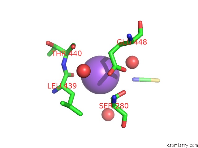
Mono view
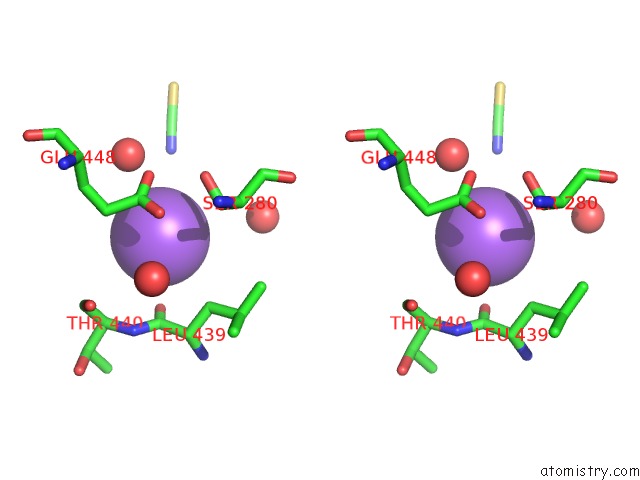
Stereo pair view
Mono view
Stereo pair view
A full contact list of Sodium with other atoms in the Na binding
site number 1 of Crystal Structure of A Glycoside Hydrolase 29 Family Member From An Unknown Rumen Bacterium within 5.0Å range:
|
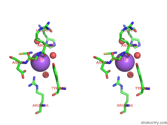
Sodium binding site 2 out of 2 in 5k9h
Go back to
Sodium binding site 2 out
of 2 in the Crystal Structure of A Glycoside Hydrolase 29 Family Member From An Unknown Rumen Bacterium

Mono view
Stereo pair view
Mono view
Stereo pair view
A full contact list of Sodium with other atoms in the Na binding
site number 2 of Crystal Structure of A Glycoside Hydrolase 29 Family Member From An Unknown Rumen Bacterium within 5.0Å range:
|
Reference:
E.L.Summers,
C.D.Moon,
R.Atua,
V.L.Arcus.
The Structure of A Glycoside Hydrolase 29 Family Member From A Rumen Bacterium Reveals Unique, Dual Carbohydrate-Binding Domains. Acta Crystallogr.,Sect.F V. 72 750 2016.
ISSN: ESSN 2053-230X
PubMed: 27710940
DOI: 10.1107/S2053230X16014072
Page generated: Mon Oct 7 22:03:27 2024
ISSN: ESSN 2053-230X
PubMed: 27710940
DOI: 10.1107/S2053230X16014072
Last articles
Zn in 9J0NZn in 9J0O
Zn in 9J0P
Zn in 9FJX
Zn in 9EKB
Zn in 9C0F
Zn in 9CAH
Zn in 9CH0
Zn in 9CH3
Zn in 9CH1