Sodium »
PDB 5jiq-5kc1 »
5k2d »
Sodium in PDB 5k2d: 1.9A Angstrom A2A Adenosine Receptor Structure with Mr Phasing Using Xfel Data
Protein crystallography data
The structure of 1.9A Angstrom A2A Adenosine Receptor Structure with Mr Phasing Using Xfel Data, PDB code: 5k2d
was solved by
A.Batyuk,
L.Galli,
A.Ishchenko,
G.W.Han,
C.Gati,
P.Popov,
M.-Y.Lee,
B.Stauch,
T.A.White,
A.Barty,
A.Aquila,
M.S.Hunter,
M.Liang,
S.Boutet,
M.Pu,
Z.-J.Liu,
G.Nelson,
D.James,
C.Li,
Y.Zhao,
J.C.H.Spence,
W.Liu,
P.Fromme,
V.Katritch,
U.Weierstall,
R.C.Stevens,
V.Cherezov,
Gpcr Network(Gpcr),
with X-Ray Crystallography technique. A brief refinement statistics is given in the table below:
| Resolution Low / High (Å) | 23.44 / 1.90 |
| Space group | C 2 2 21 |
| Cell size a, b, c (Å), α, β, γ (°) | 40.360, 180.740, 142.800, 90.00, 90.00, 90.00 |
| R / Rfree (%) | 17.4 / 20.7 |
Sodium Binding Sites:
The binding sites of Sodium atom in the 1.9A Angstrom A2A Adenosine Receptor Structure with Mr Phasing Using Xfel Data
(pdb code 5k2d). This binding sites where shown within
5.0 Angstroms radius around Sodium atom.
In total only one binding site of Sodium was determined in the 1.9A Angstrom A2A Adenosine Receptor Structure with Mr Phasing Using Xfel Data, PDB code: 5k2d:
In total only one binding site of Sodium was determined in the 1.9A Angstrom A2A Adenosine Receptor Structure with Mr Phasing Using Xfel Data, PDB code: 5k2d:
Sodium binding site 1 out of 1 in 5k2d
Go back to
Sodium binding site 1 out
of 1 in the 1.9A Angstrom A2A Adenosine Receptor Structure with Mr Phasing Using Xfel Data
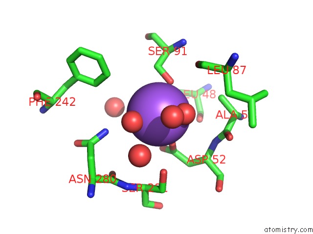
Mono view
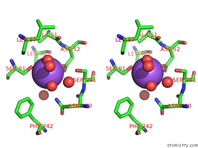
Stereo pair view
Mono view
Stereo pair view
A full contact list of Sodium with other atoms in the Na binding
site number 1 of 1.9A Angstrom A2A Adenosine Receptor Structure with Mr Phasing Using Xfel Data within 5.0Å range:
|
Reference:
A.Batyuk,
L.Galli,
A.Ishchenko,
G.W.Han,
C.Gati,
P.A.Popov,
M.Y.Lee,
B.Stauch,
T.A.White,
A.Barty,
A.Aquila,
M.S.Hunter,
M.Liang,
S.Boutet,
M.Pu,
Z.J.Liu,
G.Nelson,
D.James,
C.Li,
Y.Zhao,
J.C.Spence,
W.Liu,
P.Fromme,
V.Katritch,
U.Weierstall,
R.C.Stevens,
V.Cherezov.
Native Phasing of X-Ray Free-Electron Laser Data For A G Protein-Coupled Receptor. Sci Adv V. 2 00292 2016.
ISSN: ESSN 2375-2548
PubMed: 27679816
DOI: 10.1126/SCIADV.1600292
Page generated: Mon Oct 7 22:02:08 2024
ISSN: ESSN 2375-2548
PubMed: 27679816
DOI: 10.1126/SCIADV.1600292
Last articles
Zn in 9MJ5Zn in 9HNW
Zn in 9G0L
Zn in 9FNE
Zn in 9DZN
Zn in 9E0I
Zn in 9D32
Zn in 9DAK
Zn in 8ZXC
Zn in 8ZUF