Sodium »
PDB 5jiq-5kc1 »
5jxj »
Sodium in PDB 5jxj: Structure of the Proprotein Convertase Furin Complexed to Meta- Guanidinomethyl-Phac-Rvr-Amba in Presence of Edta
Enzymatic activity of Structure of the Proprotein Convertase Furin Complexed to Meta- Guanidinomethyl-Phac-Rvr-Amba in Presence of Edta
All present enzymatic activity of Structure of the Proprotein Convertase Furin Complexed to Meta- Guanidinomethyl-Phac-Rvr-Amba in Presence of Edta:
3.4.21.75;
3.4.21.75;
Protein crystallography data
The structure of Structure of the Proprotein Convertase Furin Complexed to Meta- Guanidinomethyl-Phac-Rvr-Amba in Presence of Edta, PDB code: 5jxj
was solved by
S.O.Dahms,
M.Arciniega,
T.Steinmetzer,
R.Huber,
M.E.Than,
with X-Ray Crystallography technique. A brief refinement statistics is given in the table below:
| Resolution Low / High (Å) | 43.23 / 2.00 |
| Space group | P 65 2 2 |
| Cell size a, b, c (Å), α, β, γ (°) | 132.080, 132.080, 155.642, 90.00, 90.00, 120.00 |
| R / Rfree (%) | 16.7 / 18.9 |
Other elements in 5jxj:
The structure of Structure of the Proprotein Convertase Furin Complexed to Meta- Guanidinomethyl-Phac-Rvr-Amba in Presence of Edta also contains other interesting chemical elements:
| Chlorine | (Cl) | 1 atom |
| Calcium | (Ca) | 1 atom |
Sodium Binding Sites:
The binding sites of Sodium atom in the Structure of the Proprotein Convertase Furin Complexed to Meta- Guanidinomethyl-Phac-Rvr-Amba in Presence of Edta
(pdb code 5jxj). This binding sites where shown within
5.0 Angstroms radius around Sodium atom.
In total 6 binding sites of Sodium where determined in the Structure of the Proprotein Convertase Furin Complexed to Meta- Guanidinomethyl-Phac-Rvr-Amba in Presence of Edta, PDB code: 5jxj:
Jump to Sodium binding site number: 1; 2; 3; 4; 5; 6;
In total 6 binding sites of Sodium where determined in the Structure of the Proprotein Convertase Furin Complexed to Meta- Guanidinomethyl-Phac-Rvr-Amba in Presence of Edta, PDB code: 5jxj:
Jump to Sodium binding site number: 1; 2; 3; 4; 5; 6;
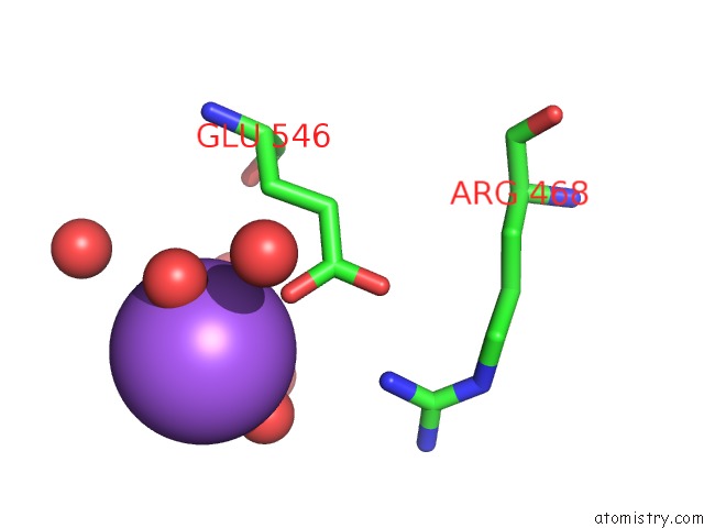
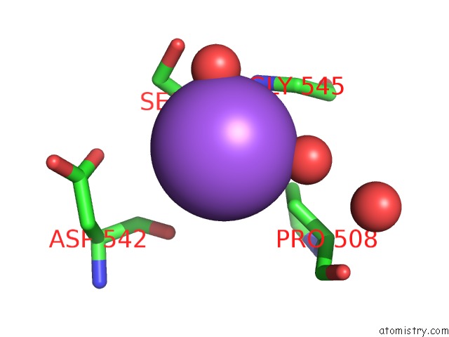
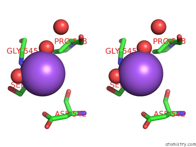
Sodium binding site 1 out of 6 in 5jxj
Go back to
Sodium binding site 1 out
of 6 in the Structure of the Proprotein Convertase Furin Complexed to Meta- Guanidinomethyl-Phac-Rvr-Amba in Presence of Edta
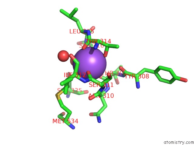
Mono view
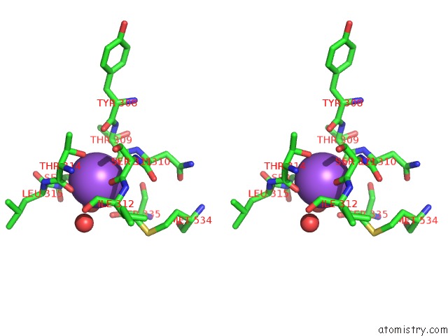
Stereo pair view
Mono view
Stereo pair view
A full contact list of Sodium with other atoms in the Na binding
site number 1 of Structure of the Proprotein Convertase Furin Complexed to Meta- Guanidinomethyl-Phac-Rvr-Amba in Presence of Edta within 5.0Å range:
|
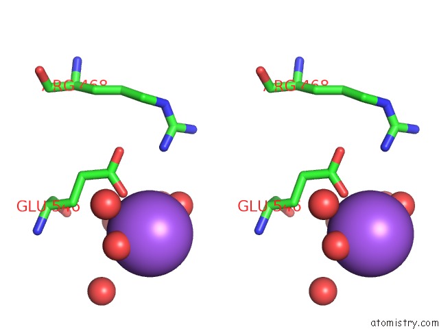
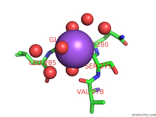
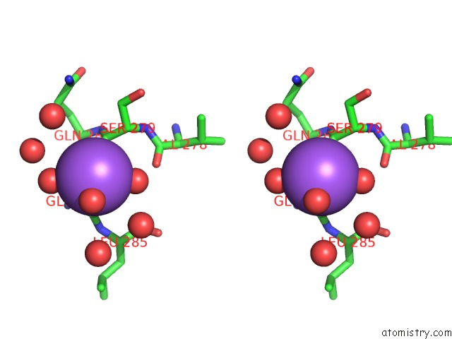
Sodium binding site 2 out of 6 in 5jxj
Go back to
Sodium binding site 2 out
of 6 in the Structure of the Proprotein Convertase Furin Complexed to Meta- Guanidinomethyl-Phac-Rvr-Amba in Presence of Edta
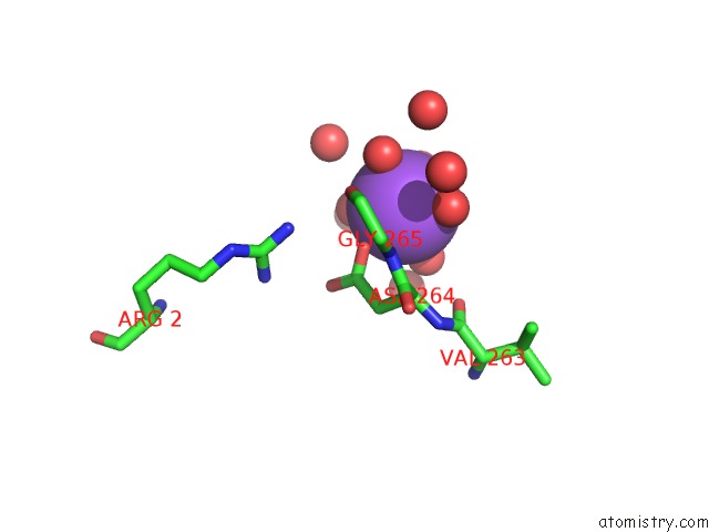
Mono view
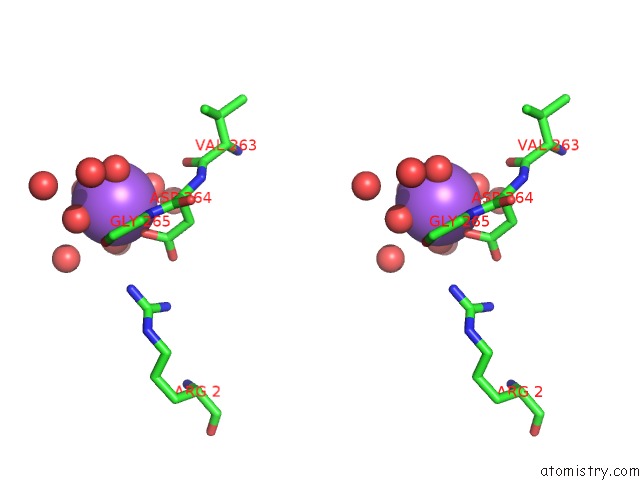
Stereo pair view
Mono view
Stereo pair view
A full contact list of Sodium with other atoms in the Na binding
site number 2 of Structure of the Proprotein Convertase Furin Complexed to Meta- Guanidinomethyl-Phac-Rvr-Amba in Presence of Edta within 5.0Å range:
|
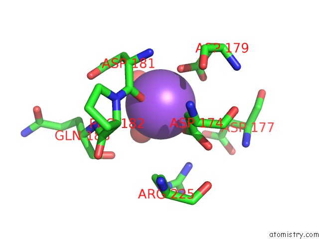
Sodium binding site 3 out of 6 in 5jxj
Go back to
Sodium binding site 3 out
of 6 in the Structure of the Proprotein Convertase Furin Complexed to Meta- Guanidinomethyl-Phac-Rvr-Amba in Presence of Edta
Mono view
Stereo pair view
Mono view
Stereo pair view
A full contact list of Sodium with other atoms in the Na binding
site number 3 of Structure of the Proprotein Convertase Furin Complexed to Meta- Guanidinomethyl-Phac-Rvr-Amba in Presence of Edta within 5.0Å range:
|
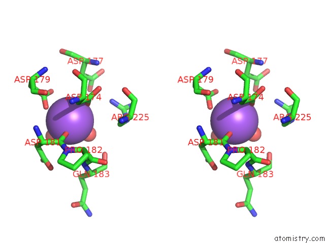
Sodium binding site 4 out of 6 in 5jxj
Go back to
Sodium binding site 4 out
of 6 in the Structure of the Proprotein Convertase Furin Complexed to Meta- Guanidinomethyl-Phac-Rvr-Amba in Presence of Edta
Mono view
Stereo pair view
Mono view
Stereo pair view
A full contact list of Sodium with other atoms in the Na binding
site number 4 of Structure of the Proprotein Convertase Furin Complexed to Meta- Guanidinomethyl-Phac-Rvr-Amba in Presence of Edta within 5.0Å range:
|
Sodium binding site 5 out of 6 in 5jxj
Go back to
Sodium binding site 5 out
of 6 in the Structure of the Proprotein Convertase Furin Complexed to Meta- Guanidinomethyl-Phac-Rvr-Amba in Presence of Edta
Mono view
Stereo pair view
Mono view
Stereo pair view
A full contact list of Sodium with other atoms in the Na binding
site number 5 of Structure of the Proprotein Convertase Furin Complexed to Meta- Guanidinomethyl-Phac-Rvr-Amba in Presence of Edta within 5.0Å range:
|
Sodium binding site 6 out of 6 in 5jxj
Go back to
Sodium binding site 6 out
of 6 in the Structure of the Proprotein Convertase Furin Complexed to Meta- Guanidinomethyl-Phac-Rvr-Amba in Presence of Edta
Mono view
Stereo pair view
Mono view
Stereo pair view
A full contact list of Sodium with other atoms in the Na binding
site number 6 of Structure of the Proprotein Convertase Furin Complexed to Meta- Guanidinomethyl-Phac-Rvr-Amba in Presence of Edta within 5.0Å range:
|
Reference:
S.O.Dahms,
M.Arciniega,
T.Steinmetzer,
R.Huber,
M.E.Than.
Structure of the Unliganded Form of the Proprotein Convertase Furin Suggests Activation By A Substrate-Induced Mechanism. Proc.Natl.Acad.Sci.Usa V. 113 11196 2016.
ISSN: ESSN 1091-6490
PubMed: 27647913
DOI: 10.1073/PNAS.1613630113
Page generated: Mon Oct 7 21:58:23 2024
ISSN: ESSN 1091-6490
PubMed: 27647913
DOI: 10.1073/PNAS.1613630113
Last articles
Zn in 9MJ5Zn in 9HNW
Zn in 9G0L
Zn in 9FNE
Zn in 9DZN
Zn in 9E0I
Zn in 9D32
Zn in 9DAK
Zn in 8ZXC
Zn in 8ZUF