Sodium »
PDB 5iaj-5imz »
5ihn »
Sodium in PDB 5ihn: Crystal Structure of the Alpha Spectrin SH3 Domain Mutant N47G
Protein crystallography data
The structure of Crystal Structure of the Alpha Spectrin SH3 Domain Mutant N47G, PDB code: 5ihn
was solved by
A.Camara-Artigas,
with X-Ray Crystallography technique. A brief refinement statistics is given in the table below:
| Resolution Low / High (Å) | 19.87 / 1.50 |
| Space group | P 21 21 21 |
| Cell size a, b, c (Å), α, β, γ (°) | 32.998, 41.863, 49.783, 90.00, 90.00, 90.00 |
| R / Rfree (%) | 16 / 17.8 |
Sodium Binding Sites:
The binding sites of Sodium atom in the Crystal Structure of the Alpha Spectrin SH3 Domain Mutant N47G
(pdb code 5ihn). This binding sites where shown within
5.0 Angstroms radius around Sodium atom.
In total 2 binding sites of Sodium where determined in the Crystal Structure of the Alpha Spectrin SH3 Domain Mutant N47G, PDB code: 5ihn:
Jump to Sodium binding site number: 1; 2;
In total 2 binding sites of Sodium where determined in the Crystal Structure of the Alpha Spectrin SH3 Domain Mutant N47G, PDB code: 5ihn:
Jump to Sodium binding site number: 1; 2;
Sodium binding site 1 out of 2 in 5ihn
Go back to
Sodium binding site 1 out
of 2 in the Crystal Structure of the Alpha Spectrin SH3 Domain Mutant N47G
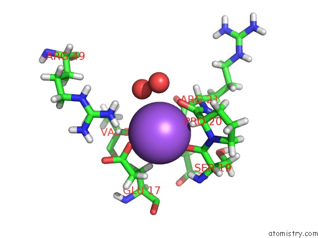
Mono view
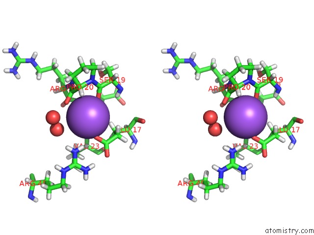
Stereo pair view
Mono view
Stereo pair view
A full contact list of Sodium with other atoms in the Na binding
site number 1 of Crystal Structure of the Alpha Spectrin SH3 Domain Mutant N47G within 5.0Å range:
|
Sodium binding site 2 out of 2 in 5ihn
Go back to
Sodium binding site 2 out
of 2 in the Crystal Structure of the Alpha Spectrin SH3 Domain Mutant N47G
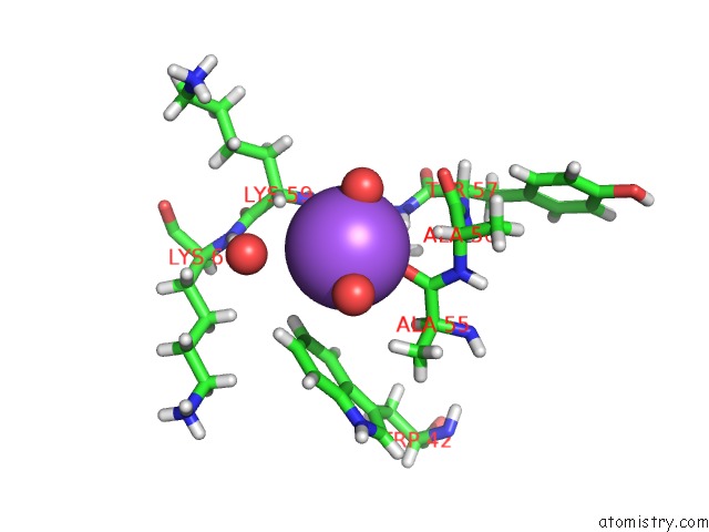
Mono view

Stereo pair view
Mono view
Stereo pair view
A full contact list of Sodium with other atoms in the Na binding
site number 2 of Crystal Structure of the Alpha Spectrin SH3 Domain Mutant N47G within 5.0Å range:
|
Reference:
A.Camara-Artigas,
A.Camara-Artigas.
N/A N/A.
Page generated: Mon Oct 7 21:35:55 2024
Last articles
Cl in 7UP4Cl in 7UOS
Cl in 7UPI
Cl in 7UMO
Cl in 7UOD
Cl in 7UN0
Cl in 7UOQ
Cl in 7UOC
Cl in 7UNO
Cl in 7UNN