Sodium »
PDB 5hn3-5i9b »
5i8f »
Sodium in PDB 5i8f: Crystal Structure of St. John'S Wort Hyp-1 Protein in Complex with Melatonin
Protein crystallography data
The structure of Crystal Structure of St. John'S Wort Hyp-1 Protein in Complex with Melatonin, PDB code: 5i8f
was solved by
J.Sliwiak,
Z.Dauter,
M.Jaskolski,
with X-Ray Crystallography technique. A brief refinement statistics is given in the table below:
| Resolution Low / High (Å) | 30.00 / 1.30 |
| Space group | C 2 2 21 |
| Cell size a, b, c (Å), α, β, γ (°) | 60.860, 89.639, 76.410, 90.00, 90.00, 90.00 |
| R / Rfree (%) | 12.8 / 15.3 |
Sodium Binding Sites:
The binding sites of Sodium atom in the Crystal Structure of St. John'S Wort Hyp-1 Protein in Complex with Melatonin
(pdb code 5i8f). This binding sites where shown within
5.0 Angstroms radius around Sodium atom.
In total 3 binding sites of Sodium where determined in the Crystal Structure of St. John'S Wort Hyp-1 Protein in Complex with Melatonin, PDB code: 5i8f:
Jump to Sodium binding site number: 1; 2; 3;
In total 3 binding sites of Sodium where determined in the Crystal Structure of St. John'S Wort Hyp-1 Protein in Complex with Melatonin, PDB code: 5i8f:
Jump to Sodium binding site number: 1; 2; 3;
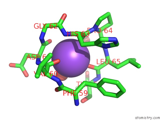
Sodium binding site 1 out of 3 in 5i8f
Go back to
Sodium binding site 1 out
of 3 in the Crystal Structure of St. John'S Wort Hyp-1 Protein in Complex with Melatonin
Mono view

Stereo pair view
Mono view
Stereo pair view
A full contact list of Sodium with other atoms in the Na binding
site number 1 of Crystal Structure of St. John'S Wort Hyp-1 Protein in Complex with Melatonin within 5.0Å range:
|
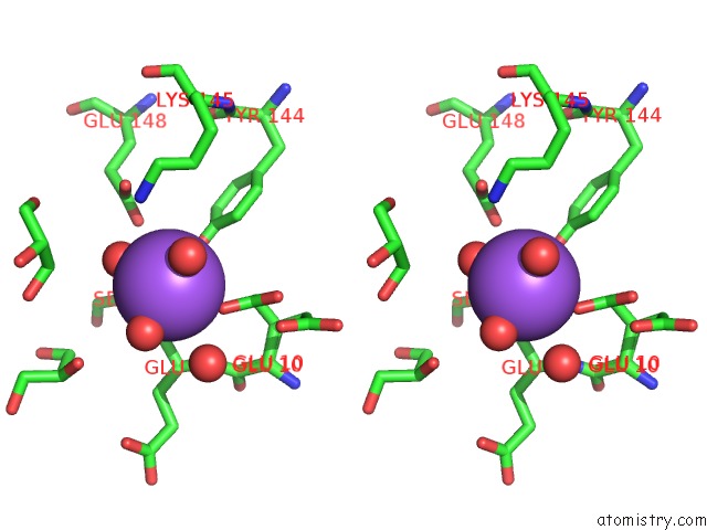
Sodium binding site 2 out of 3 in 5i8f
Go back to
Sodium binding site 2 out
of 3 in the Crystal Structure of St. John'S Wort Hyp-1 Protein in Complex with Melatonin

Mono view
Stereo pair view
Mono view
Stereo pair view
A full contact list of Sodium with other atoms in the Na binding
site number 2 of Crystal Structure of St. John'S Wort Hyp-1 Protein in Complex with Melatonin within 5.0Å range:
|
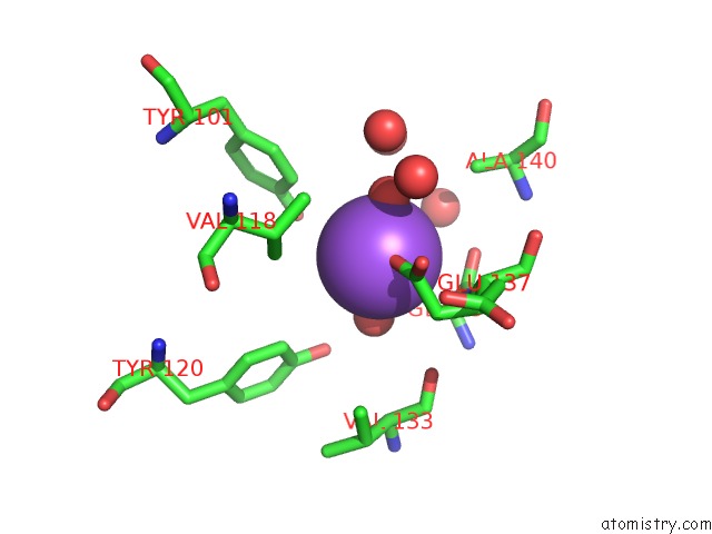
Sodium binding site 3 out of 3 in 5i8f
Go back to
Sodium binding site 3 out
of 3 in the Crystal Structure of St. John'S Wort Hyp-1 Protein in Complex with Melatonin
Mono view

Stereo pair view
Mono view
Stereo pair view
A full contact list of Sodium with other atoms in the Na binding
site number 3 of Crystal Structure of St. John'S Wort Hyp-1 Protein in Complex with Melatonin within 5.0Å range:
|
Reference:
J.Sliwiak,
Z.Dauter,
M.Jaskolski.
Crystal Structure of Hyp-1, A Hypericum Perforatum Pr-10 Protein, in Complex with Melatonin. Front Plant Sci V. 7 668 2016.
ISSN: ESSN 1664-462X
PubMed: 27242869
DOI: 10.3389/FPLS.2016.00668
Page generated: Mon Oct 7 21:33:11 2024
ISSN: ESSN 1664-462X
PubMed: 27242869
DOI: 10.3389/FPLS.2016.00668
Last articles
Zn in 9J0NZn in 9J0O
Zn in 9J0P
Zn in 9FJX
Zn in 9EKB
Zn in 9C0F
Zn in 9CAH
Zn in 9CH0
Zn in 9CH3
Zn in 9CH1