Sodium »
PDB 5hn3-5i9b »
5hy1 »
Sodium in PDB 5hy1: High Resolution Structure of Barbiturase
Enzymatic activity of High Resolution Structure of Barbiturase
All present enzymatic activity of High Resolution Structure of Barbiturase:
3.5.2.1;
3.5.2.1;
Protein crystallography data
The structure of High Resolution Structure of Barbiturase, PDB code: 5hy1
was solved by
T.S.Peat,
C.Scott,
S.Balotra,
M.Wilding,
J.Newman,
with X-Ray Crystallography technique. A brief refinement statistics is given in the table below:
| Resolution Low / High (Å) | 41.20 / 2.01 |
| Space group | I 2 2 2 |
| Cell size a, b, c (Å), α, β, γ (°) | 69.403, 82.356, 114.552, 90.00, 90.00, 90.00 |
| R / Rfree (%) | 18.6 / 22.3 |
Other elements in 5hy1:
The structure of High Resolution Structure of Barbiturase also contains other interesting chemical elements:
| Chlorine | (Cl) | 3 atoms |
Sodium Binding Sites:
The binding sites of Sodium atom in the High Resolution Structure of Barbiturase
(pdb code 5hy1). This binding sites where shown within
5.0 Angstroms radius around Sodium atom.
In total only one binding site of Sodium was determined in the High Resolution Structure of Barbiturase, PDB code: 5hy1:
In total only one binding site of Sodium was determined in the High Resolution Structure of Barbiturase, PDB code: 5hy1:
Sodium binding site 1 out of 1 in 5hy1
Go back to
Sodium binding site 1 out
of 1 in the High Resolution Structure of Barbiturase
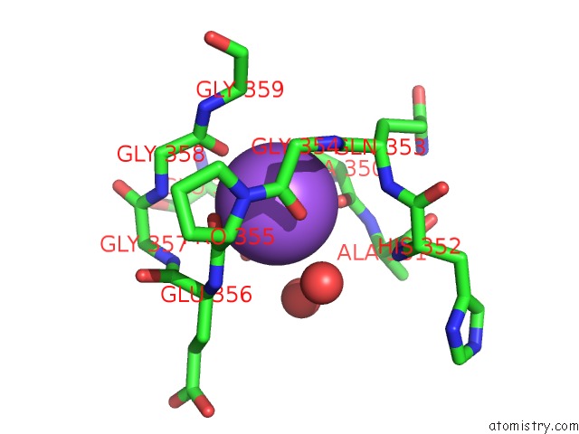
Mono view
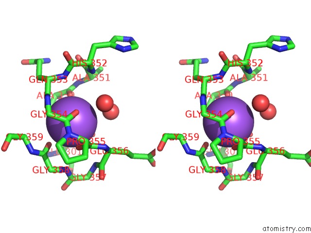
Stereo pair view
Mono view
Stereo pair view
A full contact list of Sodium with other atoms in the Na binding
site number 1 of High Resolution Structure of Barbiturase within 5.0Å range:
|
Reference:
T.S.Peat,
S.Balotra,
M.Wilding,
C.J.Hartley,
J.Newman,
C.Scott.
High-Resolution X-Ray Structures of Two Functionally Distinct Members of the Cyclic Amide Hydrolase Family of Toblerone Fold Enzymes. Appl. Environ. Microbiol. V. 83 2017.
ISSN: ESSN 1098-5336
PubMed: 28235873
DOI: 10.1128/AEM.03365-16
Page generated: Mon Oct 7 21:29:57 2024
ISSN: ESSN 1098-5336
PubMed: 28235873
DOI: 10.1128/AEM.03365-16
Last articles
Zn in 9J0NZn in 9J0O
Zn in 9J0P
Zn in 9FJX
Zn in 9EKB
Zn in 9C0F
Zn in 9CAH
Zn in 9CH0
Zn in 9CH3
Zn in 9CH1