Sodium »
PDB 5f01-5fjc »
5f7k »
Sodium in PDB 5f7k: Blood Group Antigen Binding Adhesin Baba of Helicobacter Pylori Strain 17875 in Complex with Nanobody Nb-ER19
Protein crystallography data
The structure of Blood Group Antigen Binding Adhesin Baba of Helicobacter Pylori Strain 17875 in Complex with Nanobody Nb-ER19, PDB code: 5f7k
was solved by
K.Moonens,
P.Gideonsson,
S.Subedi,
E.Romao,
S.Oscarson,
S.Muyldermans,
T.Boren,
H.Remaut,
with X-Ray Crystallography technique. A brief refinement statistics is given in the table below:
| Resolution Low / High (Å) | 58.04 / 2.17 |
| Space group | P 1 21 1 |
| Cell size a, b, c (Å), α, β, γ (°) | 50.990, 131.660, 123.460, 90.00, 94.77, 90.00 |
| R / Rfree (%) | 17.1 / 20.7 |
Sodium Binding Sites:
The binding sites of Sodium atom in the Blood Group Antigen Binding Adhesin Baba of Helicobacter Pylori Strain 17875 in Complex with Nanobody Nb-ER19
(pdb code 5f7k). This binding sites where shown within
5.0 Angstroms radius around Sodium atom.
In total only one binding site of Sodium was determined in the Blood Group Antigen Binding Adhesin Baba of Helicobacter Pylori Strain 17875 in Complex with Nanobody Nb-ER19, PDB code: 5f7k:
In total only one binding site of Sodium was determined in the Blood Group Antigen Binding Adhesin Baba of Helicobacter Pylori Strain 17875 in Complex with Nanobody Nb-ER19, PDB code: 5f7k:
Sodium binding site 1 out of 1 in 5f7k
Go back to
Sodium binding site 1 out
of 1 in the Blood Group Antigen Binding Adhesin Baba of Helicobacter Pylori Strain 17875 in Complex with Nanobody Nb-ER19
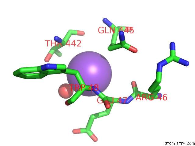
Mono view
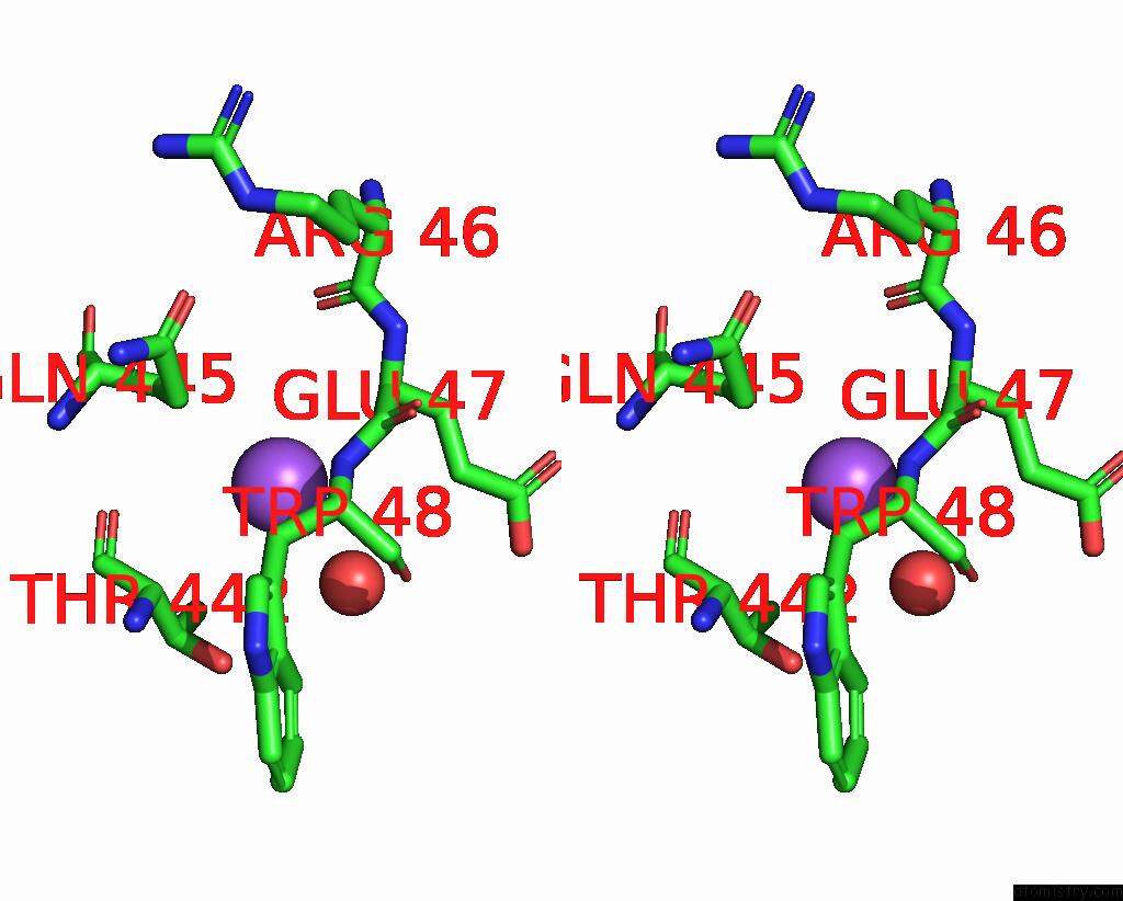
Stereo pair view
Mono view
Stereo pair view
A full contact list of Sodium with other atoms in the Na binding
site number 1 of Blood Group Antigen Binding Adhesin Baba of Helicobacter Pylori Strain 17875 in Complex with Nanobody Nb-ER19 within 5.0Å range:
|
Reference:
K.Moonens,
P.Gideonsson,
S.Subedi,
J.Bugaytsova,
E.Romao,
M.Mendez,
J.Norden,
M.Fallah,
L.Rakhimova,
A.Shevtsova,
M.Lahmann,
G.Castaldo,
K.Brannstrom,
F.Coppens,
A.W.Lo,
T.Ny,
J.V.Solnick,
G.Vandenbussche,
S.Oscarson,
L.Hammarstrom,
A.Arnqvist,
D.E.Berg,
S.Muyldermans,
T.Boren,
H.Remaut.
Structural Insights Into Polymorphic Abo Glycan Binding By Helicobacter Pylori. Cell Host Microbe V. 19 55 2016.
ISSN: ESSN 1934-6069
PubMed: 26764597
DOI: 10.1016/J.CHOM.2015.12.004
Page generated: Mon Oct 7 20:58:00 2024
ISSN: ESSN 1934-6069
PubMed: 26764597
DOI: 10.1016/J.CHOM.2015.12.004
Last articles
Zn in 9J0NZn in 9J0O
Zn in 9J0P
Zn in 9FJX
Zn in 9EKB
Zn in 9C0F
Zn in 9CAH
Zn in 9CH0
Zn in 9CH3
Zn in 9CH1