Sodium »
PDB 5ddq-5dy9 »
5djk »
Sodium in PDB 5djk: Structure of M. Tuberculosis Cysq, A Pap Phosphatase with PO4 and 2CA Bound
Enzymatic activity of Structure of M. Tuberculosis Cysq, A Pap Phosphatase with PO4 and 2CA Bound
All present enzymatic activity of Structure of M. Tuberculosis Cysq, A Pap Phosphatase with PO4 and 2CA Bound:
3.1.3.11; 3.1.3.25; 3.1.3.7;
3.1.3.11; 3.1.3.25; 3.1.3.7;
Protein crystallography data
The structure of Structure of M. Tuberculosis Cysq, A Pap Phosphatase with PO4 and 2CA Bound, PDB code: 5djk
was solved by
A.J.Fisher,
A.I.Erickson,
with X-Ray Crystallography technique. A brief refinement statistics is given in the table below:
| Resolution Low / High (Å) | 31.53 / 1.80 |
| Space group | P 21 21 21 |
| Cell size a, b, c (Å), α, β, γ (°) | 40.257, 58.225, 101.415, 90.00, 90.00, 90.00 |
| R / Rfree (%) | 15.5 / 19 |
Other elements in 5djk:
The structure of Structure of M. Tuberculosis Cysq, A Pap Phosphatase with PO4 and 2CA Bound also contains other interesting chemical elements:
| Calcium | (Ca) | 2 atoms |
Sodium Binding Sites:
The binding sites of Sodium atom in the Structure of M. Tuberculosis Cysq, A Pap Phosphatase with PO4 and 2CA Bound
(pdb code 5djk). This binding sites where shown within
5.0 Angstroms radius around Sodium atom.
In total only one binding site of Sodium was determined in the Structure of M. Tuberculosis Cysq, A Pap Phosphatase with PO4 and 2CA Bound, PDB code: 5djk:
In total only one binding site of Sodium was determined in the Structure of M. Tuberculosis Cysq, A Pap Phosphatase with PO4 and 2CA Bound, PDB code: 5djk:
Sodium binding site 1 out of 1 in 5djk
Go back to
Sodium binding site 1 out
of 1 in the Structure of M. Tuberculosis Cysq, A Pap Phosphatase with PO4 and 2CA Bound
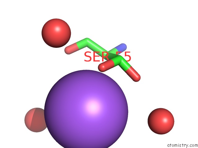
Mono view
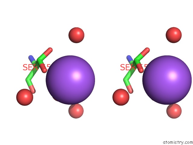
Stereo pair view
Mono view
Stereo pair view
A full contact list of Sodium with other atoms in the Na binding
site number 1 of Structure of M. Tuberculosis Cysq, A Pap Phosphatase with PO4 and 2CA Bound within 5.0Å range:
|
Reference:
A.I.Erickson,
R.D.Sarsam,
A.J.Fisher.
Crystal Structures of Mycobacterium Tuberculosis Cysq, with Substrate and Products Bound. Biochemistry V. 54 6830 2015.
ISSN: ISSN 0006-2960
PubMed: 26512869
DOI: 10.1021/ACS.BIOCHEM.5B01000
Page generated: Mon Oct 7 20:37:34 2024
ISSN: ISSN 0006-2960
PubMed: 26512869
DOI: 10.1021/ACS.BIOCHEM.5B01000
Last articles
Zn in 9J0NZn in 9J0O
Zn in 9J0P
Zn in 9FJX
Zn in 9EKB
Zn in 9C0F
Zn in 9CAH
Zn in 9CH0
Zn in 9CH3
Zn in 9CH1