Sodium »
PDB 5cd1-5cwl »
5ckq »
Sodium in PDB 5ckq: CUB1-Egf-CUB2 Domains of Rat Masp-1
Protein crystallography data
The structure of CUB1-Egf-CUB2 Domains of Rat Masp-1, PDB code: 5ckq
was solved by
R.Nan,
C.M.Furze,
D.W.Wright,
J.Gor,
R.Wallis,
S.J.Perkins,
with X-Ray Crystallography technique. A brief refinement statistics is given in the table below:
| Resolution Low / High (Å) | 76.36 / 3.70 |
| Space group | I 2 3 |
| Cell size a, b, c (Å), α, β, γ (°) | 152.710, 152.710, 152.710, 90.00, 90.00, 90.00 |
| R / Rfree (%) | 24.8 / 29.4 |
Other elements in 5ckq:
The structure of CUB1-Egf-CUB2 Domains of Rat Masp-1 also contains other interesting chemical elements:
| Calcium | (Ca) | 3 atoms |
Sodium Binding Sites:
The binding sites of Sodium atom in the CUB1-Egf-CUB2 Domains of Rat Masp-1
(pdb code 5ckq). This binding sites where shown within
5.0 Angstroms radius around Sodium atom.
In total only one binding site of Sodium was determined in the CUB1-Egf-CUB2 Domains of Rat Masp-1, PDB code: 5ckq:
In total only one binding site of Sodium was determined in the CUB1-Egf-CUB2 Domains of Rat Masp-1, PDB code: 5ckq:
Sodium binding site 1 out of 1 in 5ckq
Go back to
Sodium binding site 1 out
of 1 in the CUB1-Egf-CUB2 Domains of Rat Masp-1
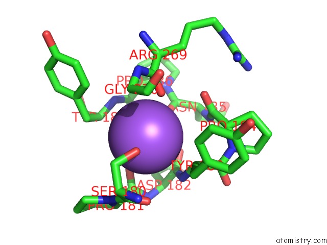
Mono view
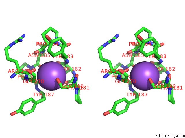
Stereo pair view
Mono view
Stereo pair view
A full contact list of Sodium with other atoms in the Na binding
site number 1 of CUB1-Egf-CUB2 Domains of Rat Masp-1 within 5.0Å range:
|
Reference:
R.Nan,
C.M.Furze,
D.W.Wright,
J.Gor,
R.Wallis,
S.J.Perkins.
Flexibility in Mannan-Binding Lectin-Associated Serine Proteases-1 and -2 Provides Insight on Lectin Pathway Activation. Structure V. 25 364 2017.
ISSN: ISSN 1878-4186
PubMed: 28111019
DOI: 10.1016/J.STR.2016.12.014
Page generated: Mon Oct 7 20:22:14 2024
ISSN: ISSN 1878-4186
PubMed: 28111019
DOI: 10.1016/J.STR.2016.12.014
Last articles
Zn in 9J0NZn in 9J0O
Zn in 9J0P
Zn in 9FJX
Zn in 9EKB
Zn in 9C0F
Zn in 9CAH
Zn in 9CH0
Zn in 9CH3
Zn in 9CH1