Sodium »
PDB 5b15-5bph »
5bjx »
Sodium in PDB 5bjx: X-Ray Structure of the Pglf 4,6-Dehydratase From Campylobacter Jejuni, Variant T395V, in Complex with Udp
Protein crystallography data
The structure of X-Ray Structure of the Pglf 4,6-Dehydratase From Campylobacter Jejuni, Variant T395V, in Complex with Udp, PDB code: 5bjx
was solved by
A.S.Riegert,
J.B.Thoden,
H.M.Holden,
with X-Ray Crystallography technique. A brief refinement statistics is given in the table below:
| Resolution Low / High (Å) | 31.00 / 1.60 |
| Space group | P 21 21 21 |
| Cell size a, b, c (Å), α, β, γ (°) | 69.229, 108.251, 108.374, 90.00, 90.00, 90.00 |
| R / Rfree (%) | 16.2 / 18.5 |
Sodium Binding Sites:
The binding sites of Sodium atom in the X-Ray Structure of the Pglf 4,6-Dehydratase From Campylobacter Jejuni, Variant T395V, in Complex with Udp
(pdb code 5bjx). This binding sites where shown within
5.0 Angstroms radius around Sodium atom.
In total 8 binding sites of Sodium where determined in the X-Ray Structure of the Pglf 4,6-Dehydratase From Campylobacter Jejuni, Variant T395V, in Complex with Udp, PDB code: 5bjx:
Jump to Sodium binding site number: 1; 2; 3; 4; 5; 6; 7; 8;
In total 8 binding sites of Sodium where determined in the X-Ray Structure of the Pglf 4,6-Dehydratase From Campylobacter Jejuni, Variant T395V, in Complex with Udp, PDB code: 5bjx:
Jump to Sodium binding site number: 1; 2; 3; 4; 5; 6; 7; 8;
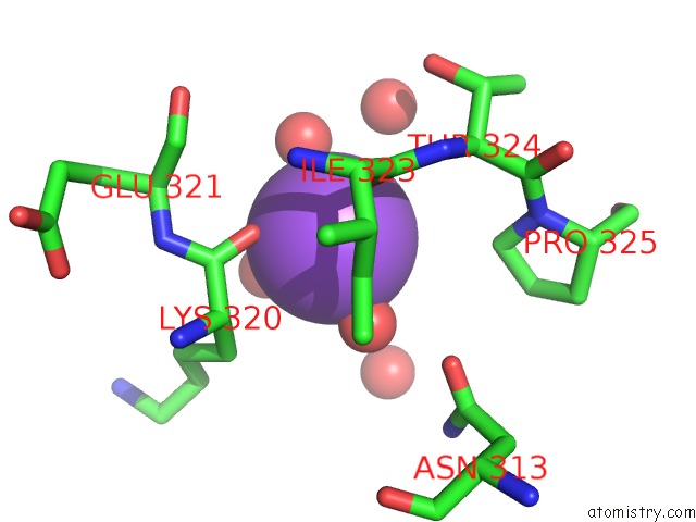
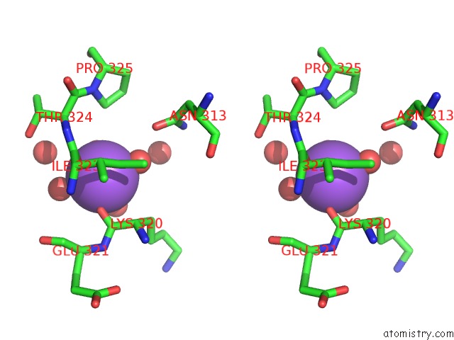
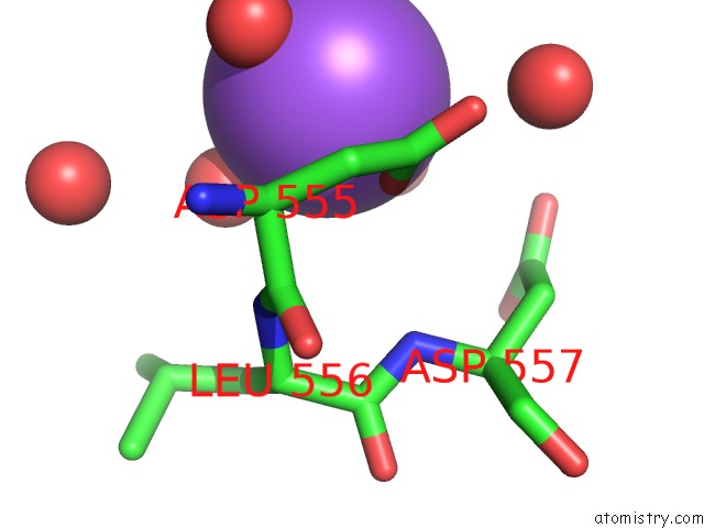
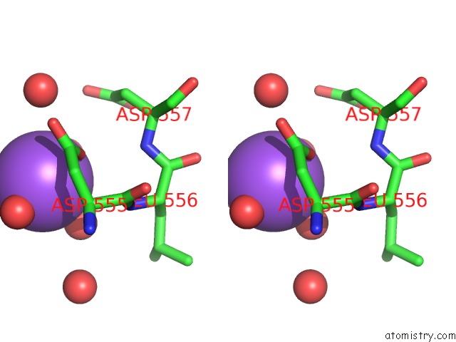
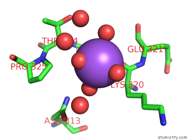
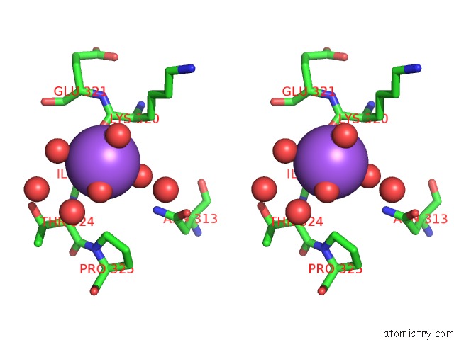
Sodium binding site 1 out of 8 in 5bjx
Go back to
Sodium binding site 1 out
of 8 in the X-Ray Structure of the Pglf 4,6-Dehydratase From Campylobacter Jejuni, Variant T395V, in Complex with Udp
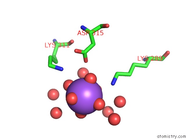
Mono view
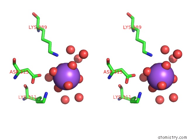
Stereo pair view
Mono view
Stereo pair view
A full contact list of Sodium with other atoms in the Na binding
site number 1 of X-Ray Structure of the Pglf 4,6-Dehydratase From Campylobacter Jejuni, Variant T395V, in Complex with Udp within 5.0Å range:
|
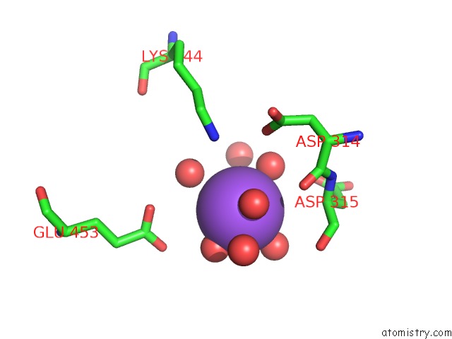
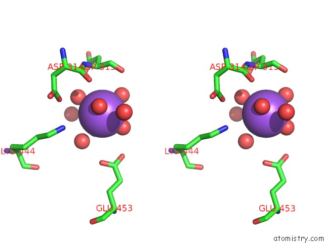
Sodium binding site 2 out of 8 in 5bjx
Go back to
Sodium binding site 2 out
of 8 in the X-Ray Structure of the Pglf 4,6-Dehydratase From Campylobacter Jejuni, Variant T395V, in Complex with Udp
Mono view
Stereo pair view
Mono view
Stereo pair view
A full contact list of Sodium with other atoms in the Na binding
site number 2 of X-Ray Structure of the Pglf 4,6-Dehydratase From Campylobacter Jejuni, Variant T395V, in Complex with Udp within 5.0Å range:
|
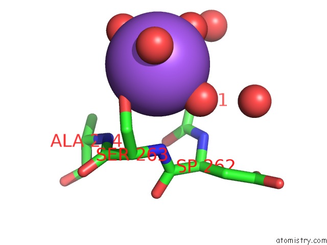
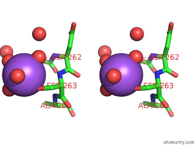
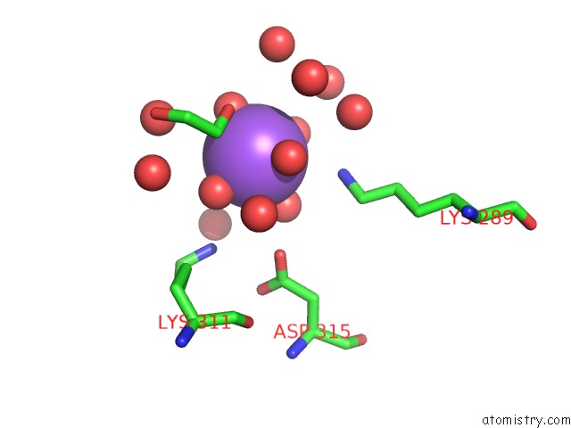
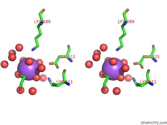
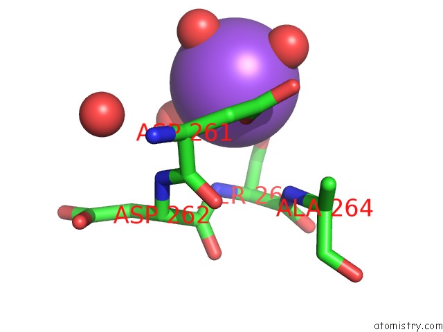
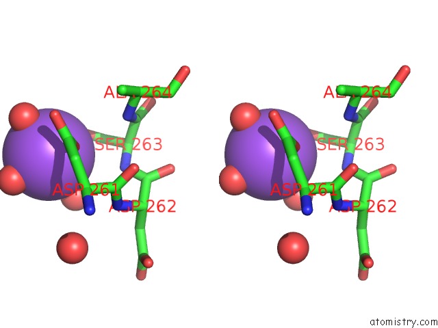
Sodium binding site 3 out of 8 in 5bjx
Go back to
Sodium binding site 3 out
of 8 in the X-Ray Structure of the Pglf 4,6-Dehydratase From Campylobacter Jejuni, Variant T395V, in Complex with Udp
Mono view
Stereo pair view
Mono view
Stereo pair view
A full contact list of Sodium with other atoms in the Na binding
site number 3 of X-Ray Structure of the Pglf 4,6-Dehydratase From Campylobacter Jejuni, Variant T395V, in Complex with Udp within 5.0Å range:
|
Sodium binding site 4 out of 8 in 5bjx
Go back to
Sodium binding site 4 out
of 8 in the X-Ray Structure of the Pglf 4,6-Dehydratase From Campylobacter Jejuni, Variant T395V, in Complex with Udp
Mono view
Stereo pair view
Mono view
Stereo pair view
A full contact list of Sodium with other atoms in the Na binding
site number 4 of X-Ray Structure of the Pglf 4,6-Dehydratase From Campylobacter Jejuni, Variant T395V, in Complex with Udp within 5.0Å range:
|
Sodium binding site 5 out of 8 in 5bjx
Go back to
Sodium binding site 5 out
of 8 in the X-Ray Structure of the Pglf 4,6-Dehydratase From Campylobacter Jejuni, Variant T395V, in Complex with Udp
Mono view
Stereo pair view
Mono view
Stereo pair view
A full contact list of Sodium with other atoms in the Na binding
site number 5 of X-Ray Structure of the Pglf 4,6-Dehydratase From Campylobacter Jejuni, Variant T395V, in Complex with Udp within 5.0Å range:
|
Sodium binding site 6 out of 8 in 5bjx
Go back to
Sodium binding site 6 out
of 8 in the X-Ray Structure of the Pglf 4,6-Dehydratase From Campylobacter Jejuni, Variant T395V, in Complex with Udp
Mono view
Stereo pair view
Mono view
Stereo pair view
A full contact list of Sodium with other atoms in the Na binding
site number 6 of X-Ray Structure of the Pglf 4,6-Dehydratase From Campylobacter Jejuni, Variant T395V, in Complex with Udp within 5.0Å range:
|
Sodium binding site 7 out of 8 in 5bjx
Go back to
Sodium binding site 7 out
of 8 in the X-Ray Structure of the Pglf 4,6-Dehydratase From Campylobacter Jejuni, Variant T395V, in Complex with Udp
Mono view
Stereo pair view
Mono view
Stereo pair view
A full contact list of Sodium with other atoms in the Na binding
site number 7 of X-Ray Structure of the Pglf 4,6-Dehydratase From Campylobacter Jejuni, Variant T395V, in Complex with Udp within 5.0Å range:
|
Sodium binding site 8 out of 8 in 5bjx
Go back to
Sodium binding site 8 out
of 8 in the X-Ray Structure of the Pglf 4,6-Dehydratase From Campylobacter Jejuni, Variant T395V, in Complex with Udp
Mono view
Stereo pair view
Mono view
Stereo pair view
A full contact list of Sodium with other atoms in the Na binding
site number 8 of X-Ray Structure of the Pglf 4,6-Dehydratase From Campylobacter Jejuni, Variant T395V, in Complex with Udp within 5.0Å range:
|
Reference:
A.S.Riegert,
J.B.Thoden,
I.C.Schoenhofen,
D.C.Watson,
N.M.Young,
P.A.Tipton,
H.M.Holden.
Structural and Biochemical Investigation of Pglf From Campylobacter Jejuni Reveals A New Mechanism For A Member of the Short Chain Dehydrogenase/Reductase Superfamily. Biochemistry V. 56 6030 2017.
ISSN: ISSN 1520-4995
PubMed: 29053280
DOI: 10.1021/ACS.BIOCHEM.7B00910
Page generated: Mon Oct 7 20:05:26 2024
ISSN: ISSN 1520-4995
PubMed: 29053280
DOI: 10.1021/ACS.BIOCHEM.7B00910
Last articles
Ca in 2ZE0Ca in 2Z8Z
Ca in 2ZDN
Ca in 2ZDM
Ca in 2ZDL
Ca in 2Z8X
Ca in 2ZDK
Ca in 2ZBJ
Ca in 2ZBD
Ca in 2ZBA